

Three-dimensional retrospective comparison of disc repositioning, condylar remodelling, and mandibular symmetry following traditional versus digital articulation techniques in patients with temporomandibular disorders
- PMID: 40887635
- PMCID: PMC12399004
- DOI: 10.1186/s12903-025-06722-8
Three-dimensional retrospective comparison of disc repositioning, condylar remodelling, and mandibular symmetry following traditional versus digital articulation techniques in patients with temporomandibular disorders
Abstract
Background: With advancements in computer technology, digital mandibular repositioning techniques are increasingly utilised in the management of temporomandibular disorders (TMDs). This study aimed to compare the differences in joint structure restoration between traditional and digital articulation techniques.
Methods: Two groups of patients with TMD (40 in each group) received traditional or digital articulation. An occlusal splint was fabricated and administered to each patient. Imaging data were collected prior to the articulation process and after the patients wore the splint for six months. Morphological and positional changes in the articular disc were analysed; condylar displacement and mandibular asymmetry were measured, and the osseous condition of the condyle was compared using three-dimensional reconstructed models.
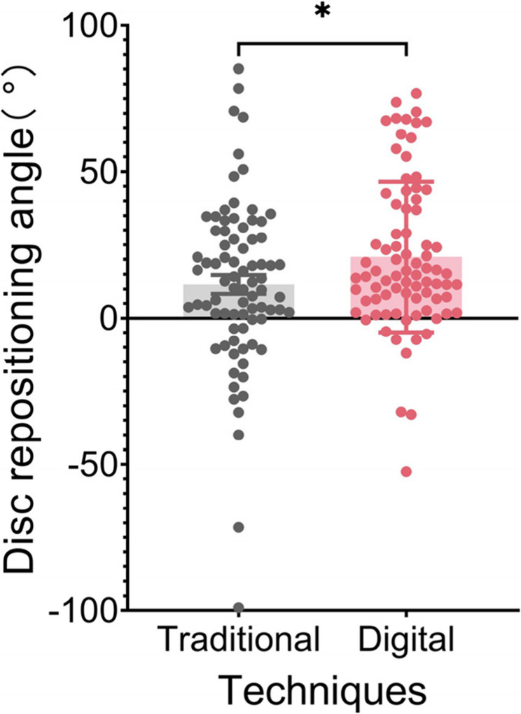
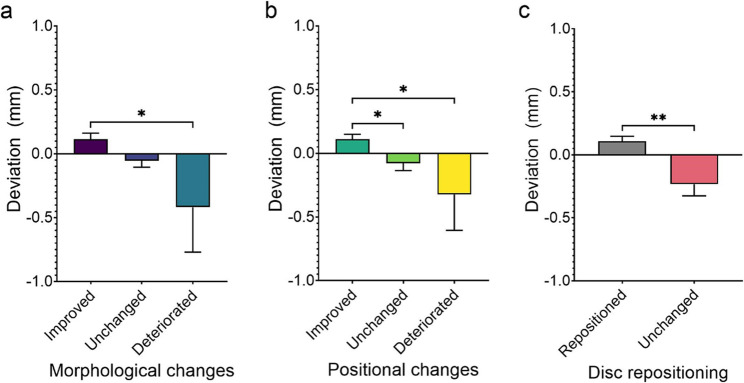
Results: The digital articulation technique performed better than the traditional approach in improving articular disc morphology (p = 0.036) and recapturing displaced discs (p = 0.032). In contrast to the traditional method [-0.1138 mm (95% CI: -0.2338, 0.0062)], digital technology notably facilitated condylar bone regeneration [0.0552 mm (95% CI: -0.0540, 0.1644)] (p = 0.040). Although no differences were observed in condylar displacement, an improvement in mandibular asymmetry was noted. A significant association was identified between articular disc repositioning and condylar bone alterations, with considerably greater bone growth noted in cases where successful articular disc repositioning was achieved [0.1072 mm (95% CI: 0.0286, 0.1858)] than in those where it was not [-0.2332 mm (95% CI: -0.4193, -0.0471)].
Conclusions: Restoring the optimal disc-condyle relationship is associated with condylar bone regeneration. The digital articulating technique offers precise control over the mandibular position and joint configuration, demonstrating satisfactory performance in improving the articular disc condition and reversing condylar bone destruction. Furthermore, it is user-friendly and highly efficient. The results suggest that the digital articulation technique may be a promising method for repositioning the articular disc and restoring condylar bone integrity.
Keywords: Articulating; Mandibular condyle; Occlusal splints; Temporomandibular disorder; Temporomandibular joint disc; Three-dimensional.
© 2025. The Author(s).
Conflict of interest statement
Declarations. Ethics approval and consent to participate: This study was approved by the Institutional Review Board of the West China Hospital of Stomatology, Sichuan University (No. WCHSIRB-OT-2019-049) and performed in line with the principles of the Declaration of Helsinki. Informed consent was obtained from all individual participants included in the study. Consent for publication: The participants/patients have provided written informed consent for the publication of their personal or clinical details and any identifiable images in this study. Competing interests: The authors declare no competing interests.
Figures


References
-
- Wright EF, Klasser GD. Manual of temporomandibular disorders. Fourth edition. Hoboken, NJ: Wiley Blackwell; 2020.
-
- Schiffman E, Ohrbach R, Truelove E, Look J, Anderson G, Goulet JP, et al. Diagnostic criteria for temporomandibular disorders (DC/TMD) for clinical and research applications: recommendations of the international RDC/TMD consortium network and orofacial pain special interest group. J Oral Facial Pain Headache. 2014;28:6–27. - PMC - PubMed
-
- Al-Moraissi EA, Farea R, Qasem KA, Al-Wadeai MS, Al-Sabahi ME, Al-Iryani GM. Effectiveness of occlusal splint therapy in the management of temporomandibular disorders: network meta-analysis of randomized controlled trials. Int J Oral Maxillofac Surg. 2020;49:1042–56. - PubMed
-
- Al-Moraissi EA, Wolford LM, Perez D, Laskin DM, Ellis E. Does orthognathic surgery cause or cure temporomandibular disorders?? A systematic review and meta-analysis. J Oral Maxillofac Surg. 2017;75:1835–47. - PubMed
Publication types
MeSH terms
Grants and funding
- 2023YFC3605600/National Key Research and Development Program of China
- 2023YFC3605600/National Key Research and Development Program of China
- 2023YFC3605600/National Key Research and Development Program of China
- 2023YFC3605600/National Key Research and Development Program of China
- 2023NSFSC0568/Natural Science Foundation of Sichuan Province
- 2023NSFSC0568/Natural Science Foundation of Sichuan Province
- 2023NSFSC0568/Natural Science Foundation of Sichuan Province
- 2023NSFSC0568/Natural Science Foundation of Sichuan Province
- LCYJ2023-YF-3/Clinical Research Project of West China Hospital of Stomatology
- LCYJ2023-YF-3/Clinical Research Project of West China Hospital of Stomatology
- LCYJ2023-YF-3/Clinical Research Project of West China Hospital of Stomatology
- LCYJ2023-YF-3/Clinical Research Project of West China Hospital of Stomatology
- RD-03-202005/R&D Program of West China Hospital of Stomatology
- RD-03-202005/R&D Program of West China Hospital of Stomatology
- RD-03-202005/R&D Program of West China Hospital of Stomatology
- RD-03-202005/R&D Program of West China Hospital of Stomatology
LinkOut - more resources
Full Text Sources
Medical
