. 2025 Aug;63(3):215-227.
doi: 10.3347/PHD.25040. Epub 2025 Aug 20.

Clonorchis sinensis dopamine transporter (CsDAT) facilitates dopamine uptake

Affiliations

Clonorchis sinensis dopamine transporter (CsDAT) facilitates dopamine uptake

Wang-Jong Lee et al. Parasites Hosts Dis. 2025 Aug.

Abstract

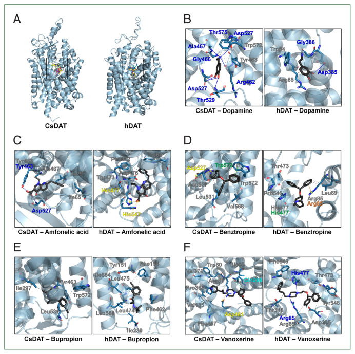
Clonorchis sinensis is a liver fluke that causes clonorchiasis, a significant public health concern in East Asia, closely associated with hepatobiliary diseases. Dopamine is an essential neurotransmitter involved in neuromuscular signaling, and its uptake by trematodes may contribute to parasite physiology and survival. This study aimed to characterize the dopamine transporter CsDAT in C. sinensis by synthesizing cDNA from adult worms and expressing it in Xenopus laevis oocytes; subsequently, uptake assays were conducted using radiolabeled dopamine. Functional assays confirmed that CsDAT mediates dopamine uptake in a sodium-dependent manner. The uptake was saturable and exhibited Michaelis-Menten kinetics with a Michaelis constant of 454.5 nM and a maximum uptake rate of 1,422.5 fmol/oocyte/h. CsDAT efficiently transported dopamine with high affinity, indicating its physiological relevance in the parasite. A 3-dimensional model of CsDAT was constructed to examine its structural features. The predicted structure contained a conserved substrate-binding pocket similar to that of other known neurotransmitter transporters. Molecular docking simulations showed that dopamine stably fits within the binding pocket. The key amino acid residues formed hydrogen bonds and hydrophobic interactions with dopamine. Interestingly, dopamine and several inhibitors demonstrated higher binding affinity to CsDAT than the human dopamine transporter. This study provides the first functional and structural insights into CsDAT. The higher inhibitor-binding affinity of CsDAT compared to human dopamine transporter suggests its potential for use in therapeutic exploration. Targeting CsDAT may facilitate the development of new therapeutic agents against clonorchiasis with minimal off-target effects on the human nervous system.

Keywords: Clonorchis sinensis; Xenopus laevis oocyte; dopamine; dopamine transporter; solute carrier family 6 member 3; uptake.


Conflict of interest statement

Conflict of interest

Jin-Hee Han serves as an editor of Parasites, Hosts and Diseases but had no involvement in the decision to publish this article. No other potential conflicts of interest relevant to this study were reported.

Figures

Fig. 1
Fig. 1
Predicted membrane topology and structural features of CsDAT. (A) Predicted membrane topology of CsDAT showing a 653-amino acid protein with 12 transmembrane helices (TM1–TM12) typical of neurotransmitter sodium symporter family transporters. Both the N- and C-termini are located in intracellular regions, and 5 potential N-glycosylation sites are identified at residues N174, N181, N189, N545, and N551. The helices are color-coded and numbered. The residue number indicates the boundaries of each helix. *, N-glycosylation site. (B) Predicted 3-dimensional structure of the CsDAT model. Transmembrane helices are color-coded and numbered. TM1, TM3, TM6, and TM8 form a central cavity that is likely involved in substrate and Na+ ion binding. The inward-facing broken helices in TM1 (1a and 1b) and TM6 (6a and 6b) may function as gates during the substrate translocation cycle. (C) Phylogenetic tree of CsDAT and related dopamine transporters from selected trematodes and mammals. CsDAT is clustered closely with the Opisthorchis felineus homolog, supporting its evolutionary conservation among parasitic flukes. The tree was constructed using the maximum-likelihood method with bootstrap values indicated at each node. Dopamine transporter sequences were obtained from the following species: Clonorchis sinensis (GAA29481), Opisthorchis felineus (TGZ63160), Paragonimus heterotremus (KAF5405469), Paragonimus westermani (KAF8563108), Fasciola hepatica (THD23603), Schistosoma haematobium (XP_035585913), Schistosoma japonicum (TNN07223), Schistosoma mekongi (KAK4475557), and Homo sapiens (AAC41720).
Fig. 2
Fig. 2
CsDAT-mediated uptake of dopamine. The uptake rates of the radiolabeled compounds were measured in water-injected (control, white bar) and CsDAT-expressing (black bar) oocytes for 1 h (mean±SE, n=8–10). The concentrations of substrates used were as follows: [3H] dopamine, 30 nM; [3H] deoxy-D-glucose, 150 nM; [3H] arginine, 100 nM; [3H] estrone sulfate, 50 nM; [3H] taurocholate, 200 nM; [14C] α-ketoglutaric acid, 5 μM; [14C] p-aminohippurate, 10 μM; and [14C] tetraethyl ammonium, 10 μM. Significant differences were observed using the Student t-test. ***P<0.001.
Fig. 3
Fig. 3
Dopamine transport properties of CsDAT. (A) The time course of [3H] dopamine uptake in CsDAT-expressing Xenopus laevis oocytes over 24–72 h after injection. (B) The time-dependent uptake of [3H] dopamine by CsDAT-expressing oocytes over incubation times ranging from 15 to 75 min. (C) Dopamine efflux assay. Oocytes were preloaded with 30 nM of [3H] dopamine for 90 min and incubated with or without excess unlabeled dopamine (1 μM and 10 μM) for 60 min to assess the substrate-induced efflux. (D) The saturation of CsDAT-mediated uptake of [3H] dopamine. The uptake rates of [3H] dopamine by the control (water-injected) or CsDAT-expressing oocytes for 60 min were measured at variable concentrations. Inset: Eadie-Hofstee plot of the concentration-dependent uptake of [3H] dopamine. V, velocity; [S], dopamine concentration. All results are presented as mean±SE (n=6–8). **P<0.01, ***P<0.001.
Fig. 4
Fig. 4
CsDAT sodium dependency. (A) Replacement of extracellular Na+ with choline or Li+ abolished dopamine uptake, demonstrating that CsDAT is sodium-dependent. (B) Sodium-binding residues within the CsDAT pocket. The sodium binding involved Gly51, Iso54, Leu399, Asp402, and Ser403 with a molecular distance of less than 3.5 Å.
Fig. 5
Fig. 5
Molecular docking analysis of dopamine and inhibitors binding to CsDAT and hDAT. (A) Overview of the binding pocket for dopamine and inhibitors, including amfonelic acid, benztropine, bupropion, and vanoxerine, in CsDAT and hDAT. (B) Detailed binding interactions of dopamine within the dopamine-binding pockets of CsDAT and hDAT. The key residues involved in hydrophobic interactions (gray) and hydrogen bonding (blue) are indicated. (C–F) Predicted binding poses and interaction residues of the dopamine transporter inhibitors amfonelic acid (C), benztropine (D), bupropion (E), and vanoxerine (F) in CsDAT and hDAT. The intewraction types are color-coded: gray, hydrophobic interactions; blue, hydrogen bonds; yellow, salt bridges; green, Pi-stacking; orange, Pi-cation interactions; cyan, halogen bonds.

References

    1. Na BK, Pak JH, Hong SJ. Clonorchis sinensis and clonorchiasis. Acta Trop. 2020;203:105309. doi: 10.1016/j.actatropica.2019.105309. - DOI - PubMed
    1. Leksomboon R, Chaijaroonkhanarak W, Arunyanart C, Umka J, Jones MK, et al. Organization of the nervous system in Opisthorchis viverrini investigated by histochemical and immunohistochemical study. Parasitol Int. 2012;61(1):107–111. doi: 10.1016/j.parint.2011.07.014. - DOI - PubMed
    1. Pax RA, Bennett JL. Neurobiology of parasitic flatworms: how much “neuro” in the biology? J Parasitol. 1992;78(2):194–205. - PubMed
    1. Terenina N, Kreshchenko N, Movsesyan S. Musculature and neurotransmitters of internal organs of trematodes (the digestive, reproductive and excretory systems) Zoology (Jena) 2022;150:125986. doi: 10.1016/j.zool.2021.125986. - DOI - PubMed
    1. Pax RA, Day TA, Miller CL, Bennett JL. Neuromuscular physiology and pharmacology of parasitic flatworms. Parasitology. 1996;113(Suppl):S83–S96. doi: 10.1017/s003118200007791x. - DOI - PubMed

MeSH terms

Substances

LinkOut - more resources