. 2025 Sep 15;152(18):dev204777.
doi: 10.1242/dev.204777. Epub 2025 Sep 18.

Mushroom bodies tiny regulates Sidekick localization to tricellular adherens junctions

Affiliations

Mushroom bodies tiny regulates Sidekick localization to tricellular adherens junctions

Dhaval Gandhi et al. Development. .

Abstract

The Drosophila cell-adhesion molecule Sidekick is a key component of tricellular adherens junctions in epithelia and localizes to specific synaptic layers in the optic lobes. Using mutagenesis of endogenous Sidekick, we showed that its enrichment at apical tricellular junctions and its function in cell rearrangement require its fifth and sixth immunoglobulin domains, but not the first four, although these mediate homophilic adhesion of mammalian Sidekick homologues. The C-terminal PDZ-binding motif of Sidekick contributes to localizing both Sidekick and its intracellular binding partner Canoe to tricellular adherens junctions. We found that the PAK4 homologue Mushroom bodies tiny also binds to the cytoplasmic domain of Sidekick. Its kinase activity is necessary for Sidekick accumulation at tricellular junctions, and over-activity mislocalizes Sidekick along bicellular junctions. However, mutating predicted Mushroom bodies tiny phosphorylation sites in Sidekick itself did not affect its localization. Sidekick localizes to the dendrites of T4 and T5 neurons independently of its extracellular and PDZ-binding domains, and of Mushroom bodies tiny. Our findings reveal an important role for a PAK4 family member in establishing specialized structures at tricellular adherens junctions.

Keywords: Junction; PAK4; Sdk; Synapse; Tricellular.


Conflict of interest statement

Competing interests The authors declare no competing or financial interests.

Figures

Figure 1:
Figure 1:. Sdk localization to tAJs does not require the first four Ig domains.
(A) Diagram of the Sdk protein showing the six Ig domains (purple), thirteen FNIII domains (yellow), transmembrane (TM) domain (gray) and PDZ-binding domain (PDZ-BD, green). Brackets indicate the extent of the deletion in each CRISPR mutant. (B-E) Mid-pupal retinas stained for Sdk (B’-E’, red in B-E), Arm (B”-E”, blue in B-E) and GFP (green) to mark clones homozygous for sdkΔIg1–4 (B), sdkΔIg1–6 (C), sdkΔIg5–6 (D) or sdkΔFN (E). Scale bars, 10 μm. (F) Quantification of Sdk levels at tAJs in clones homozygous for each mutant allele, normalized to wild-type regions of the same retinas. Error bars show mean ± s.e.m. ****, p<0.0001; ns, not significant for one-sample t test with 1 (dotted line) as the expected value. n = 10 retinas (sdkΔIg1–4), 8 (sdkΔIg1–6), 9 (sdkΔIg5–6), or 6 (sdkΔFN); each point is the mean of 10–11 junctions in that retina. Removing the first four Ig domains does not alter Sdk localization to tAJs, but deleting the fifth and sixth, all six Ig domains, or the FNIII domains strongly reduces it. Arrows in (C’, D’) indicate examples of tAJs with residual very low levels of SdkΔIg1–6 or SdkΔIg5–6.
Figure 2:
Figure 2:. Deleting the PDZ-binding motif reduces the localization of Sdk and Cno to tAJs.
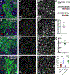
(A) Diagram of the Sdk protein indicating the extent of the ΔPDZ deletion. The C-terminal sequence is shown below with deleted amino acids colored red. (B) Mid-pupal retina stained for Sdk (B’, red in B), Arm (B”, blue in B) and GFP to mark clones homozygous for sdkΔPDZ. SdkΔPDZ localization to tAJs (yellow asterisk) is reduced relative to wild-type Sdk (blue asterisk). (C) Third instar larval wing disc stained for Cno-GFP (C’, green in C), E-cadherin (Ecad, blue) and Tomato (red) to mark clones homozygous for sdkΔPDZ. Cno-GFP localization to tAJs is reduced in sdkΔPDZ clones (yellow asterisk) relative to wild-type region (blue asterisk). Insets show enlargements of these cells, surrounded by blue (wild-type) or yellow (sdkΔPDZ) dotted lines. Scale bars, 10 μm. (D) Quantification of Sdk and Arm localization to tAJs in sdkΔPDZ clones normalized to wild-type regions of the same retinas (n=10), and of Cno-GFP localization to tAJs in sdkΔPDZ clones normalized to wild-type regions of the same wing discs (n=12). Error bars show mean ± s.e.m.. ****, p<0.0001; ns, not significant for one-sample t test with 1 (dotted line) as the expected value. Sdk and Cno localization to tAJs are partially but significantly reduced in the absence of the PDZ-binding motif of Sdk.
Figure 3:
Figure 3:. Domains required for Sdk function in tracheal development.
(A-I) show dorsolateral regions of stage 16 embryos stained for Ecad to mark AJs in the dorsal tracheal branches. (A) wild-type; (B) sdkΔ15; (C) sdkΔIg1–4; (D) sdkΔIg1–6; (E) sdkΔIg5–6; (F) sdkΔFN; (G) sdkΔPDZ; (H) sdkΔmbt; (I) mbtP1. Intercellular junctions are completely or almost completely converted to autocellular junctions, which appear as single lines, in wild-type, sdkΔIg1–4, sdkΔmbt and mbtP1 embryos at this stage. However, intercalation defects result in persistent intercellular junctions, which appear as rings, in sdk null mutants and in sdkΔIg1–6, sdkΔIg5–6, sdkΔFN and sdkΔPDZ embryos (yellow arrows), indicating that these variants have reduced function. Scale bars, 20 μm. (J) quantification of the number of tracheal branches with the categories of defects shown in the color-coded schematics on the right. Class I, wild-type; Class II, small persistent intercellular junctions; Class III, large persistent intercellular junctions; Class IV, junctions are almost entirely intercellular. Black asterisks indicate the significance with respect to control and red asterisks the significance with respect to sdkΔ15. ****, p<0.0001; **, p<0.01; ns, not significant by Kruskal-Wallis test (multiple comparisons of ranked data). n = 120 branches in 30 embryos (120/30, control), 282/36 (sdkΔ15), 71/9 (sdkΔIg1–4), 123/16 (sdkΔIg1–6), 140/18 (sdkΔIg5–6), 168/22 (sdkΔFN), 206/26 (sdkΔPDZ), 108/14 (sdkΔmbt), or 197/27 (mbtP1).
Figure 4:
Figure 4:. Sdk directly interacts with Mbt.
(A) Diagrams of the four proteins found to interact with the intracellular domain of Sdk in a yeast two-hybrid screen. The PDZ domains of Pyd, Cno and Kermit are shown in yellow, and SH3, GK, proline-rich, RA1, RA2, FHA, DIL, actin-binding, CRIB and kinase domains are also indicated. Red lines show the minimal regions of overlap between multiple clones found in the screen. (B, C) Mid-pupal retinas stained for Mbt (B’, C’, blue in B, C), Arm (B”, red in B), Ecad and N-cadherin (Cad, C”, red in C), and GFP (green) to mark sdkΔ15 null mutant clones (B) or clones overexpressing UAS-Sdk with tub-GAL4 (C). Yellow asterisks and enlarged ommatidia outlined in yellow in (B’, C’) indicate ommatidia within the clones, and blue asterisks and enlarged ommatidia outlined in blue indicate wild-type ommatidia. Scale bars, 10 μm. (D) Quantification of enrichment at cone cell tAJs normalized to bAJs for Mbt and Arm in sdk mutant clones and wild-type regions of the same retinas (n=5). (E) Quantification of Mbt and Arm intensity within lattice cells or at their bAJs in UAS-Sdk-expressing clones, normalized to wild-type regions of the same retinas (n=7). Error bars show mean ± s.e.m. **, p<0.01; *, p<0.05; ns, not significant for two-tailed t-test with Welch’s correction (D) or one-sample t test with 1 (dotted line) as the expected value (E). Sdk contributes to the enrichment of Mbt at tAJs, and Sdk overexpression mislocalizes Mbt within lattice cells.
Figure 5:
Figure 5:. Mbt kinase activity is required for Sdk localization to tAJs.
(A-D) Mid-pupal retinas stained for Sdk (A’-D’, red in A-D), Arm (A”-D”, blue in A-D) and GFP (green) to label mbtP1 clones (A), mbtP1 clones expressing UAS-mbt with tub-GAL4 (B), mbtP1 clones expressing UAS-mbtT525A with tub-GAL4 (C), or mbtP1 clones expressing UAS-mbtH19,22L with tub-GAL4 (D). Asterisks in (A”, C”, D”) show examples of mbt mutant ommatidia with abnormally arranged cells. Scale bars, 10 μm. (E) Quantification of Sdk localization at tAJs in clones normalized to wild-type regions of the same retinas. n=14 (mbtP1), 11 (mbtP1; UAS-mbt), 9 (mbtP1; UAS-mbtT525A) or 8 (mbtP1; UAS-mbtH19,21L). Error bars show mean ± s.e.m. ****, p<0.0001; ns, not significant for one-sample t test with 1 (dotted line) as the expected value. Sdk is much less enriched at tAJs in mbt mutant clones. Its enrichment is rescued by a wild-type UAS-mbt transgene, but not by UAS-mbtT525A, which has a mutation that abolishes kinase activity and may have a dominant negative effect on Sdk, or by UAS-mbtH19,22L, which has mutations that prevent Cdc42 binding.
Figure 6:
Figure 6:. Predicted phosphorylation sites in Sdk are not required for its localization by Mbt.
(A-D) Mid-pupal retinas stained for Sdk (A’-D’, red in A-D), Arm (A”-D”, blue in A-D), and GFP to label clones expressing UAS-mbtAct with tub-GAL4 (A), sdkΔIg1–6 clones expressing UAS-mbtAct with tub-GAL4 (B), sdkΔmbt clones (C), or sdkΔmbt clones expressing UAS-mbtAct with tub-GAL4 (D). Scale bars, 10 μm. (E) The two sequences in Sdk that match consensus Mbt phosphorylation sites and in which the serines predicted to be phosphorylated (S2087 and S2105) were mutated to alanines in SdkΔmbt. (F) Quantification of SdkΔIg1–6 levels at bAJs in UAS-mbtAct clones normalized to Sdk in wild-type regions of the same retinas (n=9). (G) Quantification of SdkΔmbt tAJ localization normalized to wild-type regions of the same retinas (n=9). (H) Quantification of Sdk or SdkΔmbt localization at bAJs relative to tAJs in the presence or absence of activated Mbt. n=18 (wild-type), 13 (mbtAct), 8 (sdkΔmbt) or 10 (sdkΔmbt; mbtAct). Error bars show mean ± s.e.m. ****, p<0.0001; ***, p<0.001; ns, not significant for one-sample t test with 1 (dotted line) as the expected value (F, G) or unpaired two-tailed t test with Welch’s correction (H). Activated Mbt mislocalizes wild-type Sdk along bicellular junctions, and also increases the localization of Sdk lacking its Ig domains to bAJs. Mutating two predicted phosphorylation sites in the intracellular domain of Sdk does not alter its localization to tAJs or prevent it from being mislocalized by activated Mbt. Asterisks in (A’, B’, D’) mark examples of bAJs with increased levels of wild-type or mutant Sdk proteins.
Figure 7:
Figure 7:. Localization of Sdk to dendrites does not require the same mechanism as localization to tAJs.
(A-J) show 72 h APF pupal brains stained for Sdk (green in A-G, I, J) or Mbt (green in H) and Elav (magenta in A-D, H), Ncad (blue in E, F, magenta in G, I J) or Pyd (E’, F’, red in E, F). (A, E) w1118 control; (B) sdklinker; (C) sdkΔIg1–4; (D) sdkΔIg1–6; (F) sdkΔPDZ; (G) sdkΔFN; (H) FRT19A control; (I) mbtP1; (J) T4/5-GAL4, UAS-mbtAct. Sdk is localized to the medulla layers M3 and M10 and the lobula layer Lo1 in all genotypes. Pyd overlaps with Sdk in M10 and Lo1, but is not affected in sdkΔPDZ. Mbt is also localized to M10 and Lo1, but loss or gain of Mbt activity does not affect Sdk localization. Scale bars, 20 μm. (K) Model for Sdk localization to tAJs. Ig domains 5 and 6, but not domains 1–4, are necessary for Sdk accumulation at tAJs, and the FNIII domains are important for export to the plasma membrane but also contribute to tAJ localization. Mbt directly binds to Sdk and may control its localization by phosphorylating Sdk, its binding partners, or other proteins that alter cell bond tension. (L) Model for Sdk localization to synapses. Changes in the Sdk protein do not affect its localization to synapses, suggesting that sdk mRNA may be transported there. Sdk localization to the tips of T5 dendritic arbors, where Tm9 forms synapses, might require homophilic adhesion or Mbt activity.

References

    1. Astigarraga S, Douthit J, Tarnogorska D, Creamer MS, Mano O, Clark DA, Meinertzhagen IA and Treisman JE (2018). Drosophila Sidekick is required in developing photoreceptors to enable visual motion detection. Development 145, dev158246. - PMC - PubMed
    1. Baskaran Y, Tay FP, Ng EYW, Swa CLF, Wee S, Gunaratne J and Manser E (2021). Proximity proteomics identifies PAK4 as a component of Afadin-Nectin junctions. Nat Commun 12, 5315. - PMC - PubMed
    1. Bischof J, Maeda RK, Hediger M, Karch F and Basler K (2007). An optimized transgenesis system for Drosophila using germ-line-specific phiC31 integrases. Proc Natl Acad Sci U S A 104, 3312–3317. - PMC - PubMed
    1. Borst A, Haag J and Mauss AS (2020). How fly neurons compute the direction of visual motion. J Comp Physiol A Neuroethol Sens Neural Behav Physiol 206, 109–124. - PMC - PubMed
    1. Byri S, Misra T, Syed ZA, Batz T, Shah J, Boril L, Glashauser J, Aegerter-Wilmsen T, Matzat T, Moussian B, et al. (2015). The triple-repeat protein Anakonda controls epithelial tricellular junction formation in Drosophila. Dev Cell 33, 535–548. - PubMed

Substances