. 2025 Sep;29(17):e70683.
doi: 10.1111/jcmm.70683.

Butyrate Reducing Bone Mass Loss by Regulating the Expression of m6A Methyltransferase METTL3 in Implant-Associated Staphylococcus aureus Osteomyelitis

Affiliations

Butyrate Reducing Bone Mass Loss by Regulating the Expression of m6A Methyltransferase METTL3 in Implant-Associated Staphylococcus aureus Osteomyelitis

Chongkai Sun et al. J Cell Mol Med. 2025 Sep.

Abstract

Staphylococcus aureus (S. aureus) has been identified as a hindrance to osteoblast differentiation, thereby contributing significantly to the development of osteomyelitis. Consequently, exploring pharmaceutical interventions targeting osteoblast differentiation mediated by S. aureus may present a novel approach for treating osteomyelitis. It has been reported that N6-methyladenosine (m6A) methylation is highly associated with infection. Moreover, studies continue to validate that short-chain fatty acids play an important role in transcriptional modification and have been considered as a potential treatment for S. aureus infection. Our research aimed to examine the impact and underlying mechanism of butyrate, a short-chain fatty acid, in reducing the inhibitory influence exerted by S. aureus on osteoblast differentiation. The concentration of butyrate beneficial to MC3T3-E1 cell viability was screened by Cell Counting Kit-8 (CCK8) assay. Reverse transcription-quantitative PCR (RT-qPCR), Western blotting, and alkaline phosphatase (ALP)staining were used to verify the osteogenic indexes and the expression levels of m6A methylation-related proteins in MC3T3-E1 cells infected with S. aureus at different time points. Besides, the same methods were used to verify the effects of butyrate stimulation and METTL3 knockdown on the osteogenic ability of MC3T3-E1 cells. H&E staining, Goldner staining, and immunohistochemical staining were used to verify the effect of butyrate on mice with endo-plant associated S. aureus osteomyelitis. The potential mechanisms of METTL3 and autophagy in MC3T3-E1 cells were studied by Western blotting. In vitro experiments, we found that butyrate significantly enhanced the expression of osteogenic-related genes down-regulated by infection in MC3T3-E1 cells induced by S. aureus, including RUNX2, OCN, and ALP. In addition, METTL3, an important m6A methyltransferase, was significantly up-regulated in S. aureus-infected MC3T3-E1 cells, but its expression could be down-regulated by butyrate. Inhibiting the expression of METTL3 by siRNA could effectively rescue the osteogenic markers down-regulated by S. aureus infection in MC3T3-E1 cells, which was similar to the results of butyrate treatment. In vivo experiments had shown that butyrate could alleviate inflammation and osteogenic activity in implant-associated osteomyelitis. The construction of bone marrow METTL3 low-expression mice using siRNA also showed similar effects on osteogenic activity. Additionally, Western blotting confirmed that knocking down METTL3 rescued the autophagy imbalance caused by S. aureus infection in MC3T3-E1 cells. In general, our research demonstrated that butyrate effectively alleviated the hindrance of osteoblast activity induced by S. aureus infection by suppressing the expression of METTL3, suggesting that butyrate may be a novel treatment for S. aureus osteomyelitis.

Keywords: Staphylococcus aureus; METTL3; N6‐methyladenosine; butyrate; osteomyelitis.


Conflict of interest statement

The authors declare no conflicts of interest.

Figures

FIGURE 1
FIGURE 1
The effect of butyrate on the viability of MC3T3‐E1 cells and the inhibition of inflammation induced by S. aureus . CCK8 assay was used to determine the effect of butyrate on the viability of MC3T3‐E1 cells and S. aureus treated MC3T3‐E1 cells at 24, 48, 72 h and 7 days. n = 3 in each group. *p < 0.05, **p < 0.01, ***p < 0.001 versus 0 μg/mL. # p < 0.05, ## p < 0.01, ### p < 0.001 versus 10 μg/mL. ^p < 0.05, ^^p < 0.01, ^^^p < 0.001 versus 20 μg/mL. + p < 0.05, ++ p < 0.01, +++ p < 0.001 versus 50 μg/mL. & p < 0.05, && p < 0.01, &&& p < 0.001 versus 75 μg/mL. $ p < 0.05, $$ p < 0.01, $$$ p < 0.001 versus 100 μg/mL. % p < 0.05, %% p < 0.01, %%% p < 0.001 versus 200 μg/mL. S. aureus , Staphylococcus aureus .
FIGURE 2
FIGURE 2
A certain concentration of butyrate could increase the down‐regulated osteogenic capability (A) The protein levels of osteogenic indexes (RUNX2, OCN) were analysed by Western blotting after 0, 10 and 20 μg/mL butyrate treatment with or without S. aureus infection for 24 h. (B) The protein levels of osteogenic indexes (RUNX2, OCN) were analysed by Western blotting after 0,10 and 20 μg/mL butyrate treatment with or without S. aureus infection for 7 days. (C, D) Protein quantification of (B) by ImageJ. n = 3 in each group. (E–G) qPCR analysis of osteogenic indexes (RUNX2, OCN and ALP) 7 days after 0,10 and 20 μg/mL butyrate treatment with or without S. aureus infection. (H) Scanning and microscopic images of MC3T3‐E1 cells treated with and without S. aureus infection at 10 and 20 μg/mL butyrate for 7 days after ALP staining. *p < 0.05, **p < 0.01, ***p < 0.001 versus Ctrl. # p < 0.05, ## p < 0.01, ### p < 0.001 versus OM. ^p < 0.05, ^^p < 0.01, ^^^p < 0.001 versus OM +  S. aureus .+ p < 0.05,++ p < 0.01,+++ p < 0.001 versus Butyate 10. & p < 0.05, && p < 0.01, &&& p < 0.001 versus Butyrate 10 +  S. aureus . % p < 0.05,%% p < 0.01,%%% p < 0.001 versus Butyrate 20. S. aureus , Staphylococcus aureus; RUNX2, Runt‐related transcription factor 2; OCN, osteocalcin; ALP, Alkaline Phosphatase; OM, Osteogenic medium; qPCR, quantitative PCR.
FIGURE 3
FIGURE 3
After 7 days of S. aureus infection, METTL3 levels were up‐regulated in MC3T3‐E1 cells, which could be reversed by low concentration butyrate treatment. (A) The protein levels of m6A methylation‐related genes (METTL3, METTL14, FTO and ALKBH5) in MC3T3‐E1 cells treated with or without S. aureus infection in osteogenic medium for 24 h or 7 days were analysed by Western blotting. (B) Protein quantification of (A) by ImageJ. n = 3 in each group. *p < 0.05, **p < 0.01 versus Ctrl. (C) qPCR analysis of m6A methyltransferase METTL3 in MC3T3‐E1 cells treated with or without S. aureus infection and with or without osteogenic medium for 24 h or 7 days. *p < 0.05 versus Ctrl. (D) The protein levels of m6A methylation‐related genes (METTL3, METTL14, FTO, ALKBH5) in MC3T3‐E1 cells treated with 0, 10 or 20 μg/mL butyrate groups with or without S. aureus infection for 24 h were analysed by Western blotting. (E) The protein levels of m6A methylation‐related genes (METTL3, METTL14, FTO, ALKBH5) in MC3T3‐E1 cells treated with 0, 10 or 20 μg/mL butyrate groups with or without S. aureus infection for 7 days were analysed by Western blotting. (F) Protein quantification of (E) by ImageJ. n = 3 in each group. (G) qPCR analysis of m6A methyltransferase METTL3 in MC3T3‐E1 cells treated with 0, 10 or 20 μg/mL butyrate with or without S. aureus infection for 7 days. *p < 0.05, **p < 0.01, ***p < 0.001 versus Ctrl. # p < 0.05, ## p < 0.01, ### p < 0.001 versus OM. ^p < 0.05, ^^p < 0.01, ^^^p < 0.001 versus OM +  S. aureus . + p < 0.05, ++ p < 0.01, +++ p < 0.001 versus Butyate 10. & p < 0.05, && p < 0.01, &&& p < 0.001 versus Butyrate 10 +  S. aureus . % p < 0.05, %% p < 0.01, %%% p < 0.001 versus Butyrate 20. S. aureus , Staphylococcus aureus ; OM, Osteogenic media; m6A, N6 methyladenosine; METTL3, methyltransferase‐like 3; METTL14, methyltransferase‐like 14; ALKBH5, AlkB homologue 5; FTO, Fat mass and obesity associated gene; qPCR, quantitative PCR.
FIGURE 4
FIGURE 4
Screening of siMETTL3 (A) CCK8 assay was used to determine the effect of siMETTL3 on the viability of MC3T3‐E1 cells. (B) The protein level of m6A methyltransferase METTL3 in MC3T3‐E1 cells treated with three different siMETTL3 and siRNA were analysed by Western blotting. (C) Protein quantification of (B) by ImageJ. n = 3 in each group. (D) qPCR analysis of m6A methyltransferase METTL3 of MC3T3‐E1 cells treated with three different siMETTL3 or siRNA. *p < 0.05, **p < 0.01 versus Ctrl. ## p < 0.01 versus siRNA. ^p < 0.05 versus siMETTL3‐+1. p < 0.05 versus siMETTL3‐2. siRNA, small interfering RNA; si‐METTL3, siRNA targeting METTL3; METTL3, methyltransferase‐like 3; qPCR, quantitative PCR.
FIGURE 5
FIGURE 5
Silencing METTL3 restored the osteogenic index down‐regulated by S. aureus infection in MC3T3‐E1 cells. (A) The protein levels of m6A methyltransferase METTL3, osteogenic index RUNX2 and OCN in MC3T3‐E1 cells treated with or without S. aureus , siMETTL3, siRNA and butyrate were analysed by Western blotting. (B–D) Protein quantification of (A) by ImageJ. n = 3 in each group. (E–H) qPCR analysis of m6A methyltransferase METTL3, osteogenic index RUNX2, OCN and ALP of MC3T3‐E1 cells treated with or without S. aureus , siMETTL3, siRNA and butyrate for 7 days. n = 3 in each group. (I) Scanning and microscopic images of MC3T3‐E1 cells treated with or without S. aureus , siMETTL3, siRNA and butyrate for 7 days after ALP staining. *p < 0.05, **p < 0.01, ***p < 0.001 versus Ctrl. # p < 0.05, ## p < 0.01, ### p < 0.001 versus siRNA. ^p < 0.05, ^^p < 0.01, ^^^p < 0.001 versus siMETTL3. + p < 0.05, ++ p < 0.01, +++ p < 0.001 versus Ctrl + S. aureus. & p < 0.05, && p < 0.01, &&& p < 0.001 versus siRNA+ S. aureus . % p < 0.05, %% p < 0.01, %%% p < 0.001 versus siMETTL3 +  S. aureus . siRNA, small interfering RNA; si‐METTL3, siRNA targeting METTL3; METTL3, methyltransferase‐like 3; S. aureus , Staphylococcus aureus; m6A, N6‐methyladenosine; RUNX2, Runt‐related transcription factor 2; OCN, osteocalcin; ALP, alkaline phosphatase; qPCR, quantitative PCR.
FIGURE 6
FIGURE 6
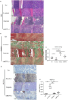
Mouse femur H&E staining, Goldner trichrome staining results, and METTL3 immunohistochemistry results revealed the anti‐inflammatory, bone promoting, and METTL3 inhibiting effects of butyrate in a plant associated Staphylococcus aureus osteomyelitis mouse model (A) H&E staining was performed on the bones of Ctrl group, S. aureus group, butyrate group and siMETTL3 group. The scale represents 200, 100 and 50 μm from left to right, respectively. (B) Goldner staining was performed on the bones of Ctrl group, S. aureus group, butyrate group and siMETTL3 group. The scale represents 200, 100 and 50 μm from left to right, respectively. (C) The relative area ratio of new bone to mature bone was analysed by Image Pro Plus 6.0 for (B). n = 6 in each group. *p < 0.05, **p < 0.01. (D) Representative images of METTL3 immunohistochemical staining of bone in Ctrl group, S. aureus group, butyrate group and siMETTL3 group. The scale from left to right represents 50 and 20 μm, respectively. (E) Quantitative analysis of METTL3 was performed by Image Pro Plus 6.0. The immunohistochemical staining average optical density value (MOD) of immunohistochemical staining was used for quantitative analysis of (D). n = 6 in each group. *p < 0.05, **p < 0.01, ***p < 0.001. si‐METTL3, siRNA targeting METTL3; S. aureus , Staphylococcus aureus ; METTL3, methyltransferase‐like 3.
FIGURE 7
FIGURE 7
Immunohistochemical results of RUNX2, OCN, CD31 and Angiogenin in the femur of mice (A–D) Representative images of RUNX2, OCN, CD31 and Angiogenin immunohistochemical staining of bone in the Ctrl group, S. aureus group, Butyrate group and siMETTL3 group. The scale from left to right represents 50 and 20 μm, respectively. (E–H) Quantitative analysis of RUNX2, OCN, CD31 and Angiogenin was performed by Image Pro Plus 6.0. The immunohistochemical staining average optical density value (MOD) of immunohistochemical staining was used for quantitative analysis of (A–D). n = 6 in each group. *p < 0.05, **p < 0.01, ***p < 0.001. si‐METTL3, siRNA targeting METTL3; S. aureus , Staphylococcus aureus ; RUNX2, Runt‐related transcription factor 2; OCN, osteocalcin.
FIGURE 8
FIGURE 8
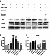
Silent METTL3 can save autophagy of MC3T3‐E1 cells damaged by S. aureus infection (A) The protein levels of P62 and LC3B in MC3T3‐E1 cells treated with or without S. aureus , siMETTL3, siRNA and butyrate for 7 days were analysed by Western blotting. (B) (C) Protein quantification of (A) by ImageJ. n = 3 in each group. *p < 0.05, **p < 0.01, ***p < 0.001 versus Ctrl. # p < 0.05, ## p < 0.01, ### p < 0.001 versus siRNA. ^p < 0.05, ^^p < 0.01, ^^^p < 0.001 versus +siMETTL3. p < 0.05, ++ p < 0.01, +++ p < 0.001 versus Ctrl+S. aureus. & p < 0.05, && p < 0.01, &&& p < 0.001 versus siRNA+ S. aureus . % p < 0.05, %% p < 0.01, %%% p < 0.001 versus siMETTL3 +  S. aureus . siRNA, small interfering RNA; si‐METTL3, siRNA targeting METTL3; S. aureus . Staphylococcus aureus.

Similar articles

References

    1. Kavanagh N., Ryan E. J., Widaa A., et al., “Staphylococcal Osteomyelitis: Disease Progression, Treatment Challenges, and Future Directions,” Clinical Microbiology Reviews 31 (2018): 10‐1128. - PMC - PubMed
    1. Panteli M. and Giannoudis P. V., “Chronic Osteomyelitis: What the Surgeon Needs to Know,” EFORT Open Reviews 1 (2016): 128–135. - PMC - PubMed
    1. Kremers H. M., Nwojo M. E., Ransom J. E., Wood‐Wentz C. M., L. J. Melton, 3rd , and P. M. Huddleston, 3rd , “Trends in the Epidemiology of Osteomyelitis: A Population‐Based Study, 1969 to 2009,” Journal of Bone and Joint Surgery. American Volume 97 (2015): 837–845. - PMC - PubMed
    1. Tong S. Y., Davis J. S., Eichenberger E., et al., “ <styled-content style="fixed-case"> Staphylococcus aureus </styled-content> Infections: Epidemiology, Pathophysiology, Clinical Manifestations, and Management,” Clinical Microbiology Reviews 28 (2015): 603–661. - PMC - PubMed
    1. Wu W. J., Xia C. L., Ou S. J., et al., “Prophylactic Effects of NFκB Essential Modulator‐Binding Domain Peptides on Bone Infection: An Experimental Study in a Rabbit Model,” Journal of Inflammation Research 15 (2022): 2745–2759. - PMC - PubMed

MeSH terms

LinkOut - more resources