

Correlation between balanced fast field echo sequence and intraoperative findings in the surgical treatment of an intradural spinal arachnoid cyst: illustrative case
- PMID: 40889395
- PMCID: PMC12400851
- DOI: 10.3171/CASE25145
Correlation between balanced fast field echo sequence and intraoperative findings in the surgical treatment of an intradural spinal arachnoid cyst: illustrative case
Abstract
Background: Spinal arachnoid cysts can cause myelopathy through spinal cord compression. While MRI is the standard for diagnosis, traditional sequences may not clearly define cyst borders and septations, which are important for guiding surgical intervention. Balanced fast field echo (B-FFE) is an MRI sequence that highlights small arachnoid membranes within and at the borders of CSF spaces.
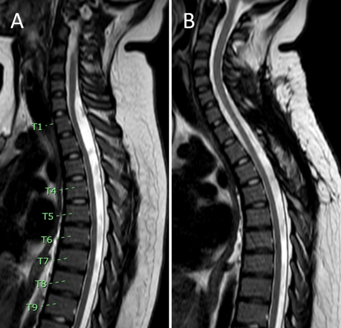
Observations: The authors report the case of a 13-year-old female who presented with progressive lower extremity paresthesias and weakness and urinary incontinence. MRI revealed an intradural cervicothoracic arachnoid cyst (C7-T3) dorsal to the spinal cord. B-FFE was used to identify the upper and lower borders of the intradural arachnoid cyst and its internal septations. These findings corresponded precisely with intraoperative findings and guided fenestration at the cyst's cranial, caudal, and internal septal ends. Postoperatively, the patient's symptoms resolved, and MRI confirmed the resolution of mass effect. At the 10- and 30-month follow-ups, there was no evidence of cyst recurrence clinically or radiographically.
Lessons: The authors raise awareness of the clinical utility of B-FFE imaging for intradural spinal arachnoid cysts. Due to its ability to demonstrate cyst borders and internal septations, it offers an alternative to more invasive tests, especially in the pediatric population. https://thejns.org/doi/10.3171/CASE25145.
Keywords: B-FFE; balanced fast field echo; balanced steady-state free precession; magnetic resonance imaging; pediatric neuroimaging; spinal arachnoid cyst.
Figures


References
-
- Bond AE Zada G Bowen I McComb JG Krieger MD.. Spinal arachnoid cysts in the pediatric population: report of 31 cases and a review of the literature. J Neurosurg Pediatr. 2012;9(4):432-441. - PubMed
-
- Hughes G Ugokwe K Benzel EC.. A review of spinal arachnoid cysts. Cleve Clin J Med. 2008;75(4):311-315. - PubMed
-
- Patel DM Weinberg BD Hoch MJ. CT.. CT myelography: clinical indications and imaging findings. Radiographics. 2020;40(2):470-484. - PubMed
-
- Nabors MW, Pait TG, Byrd EB.Updated assessment and current classification of spinal meningeal cysts. J Neurosurg. 1988;68(3):366-377. - PubMed
-
- Koh JS Chang UK Kim SH Haddix T. Chapter 5.. Intradural extramedullary cystic lesions. In: Kim DH, Chang U-K, Kim S-H, Bilsky MH, eds. Tumors of the Spine. W.B. Saunders; 2008:71-80.
LinkOut - more resources
Full Text Sources
Miscellaneous
