. 2025 Sep 1;16(1):667.
doi: 10.1038/s41419-025-07991-5.

MAP3K13-232aa encoded by circMAP3K13 enhances cisplatin-induced pyroptosis by directly binding to IKKα in gastric adenocarcinoma

Affiliations

MAP3K13-232aa encoded by circMAP3K13 enhances cisplatin-induced pyroptosis by directly binding to IKKα in gastric adenocarcinoma

Kaining Du et al. Cell Death Dis. .

Abstract

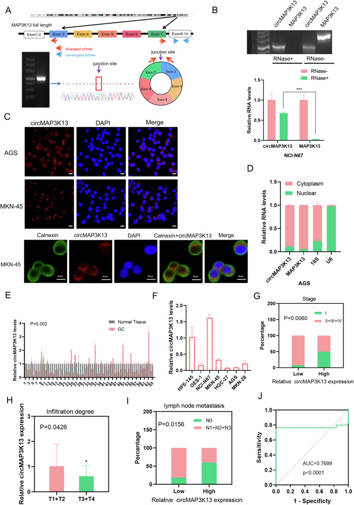
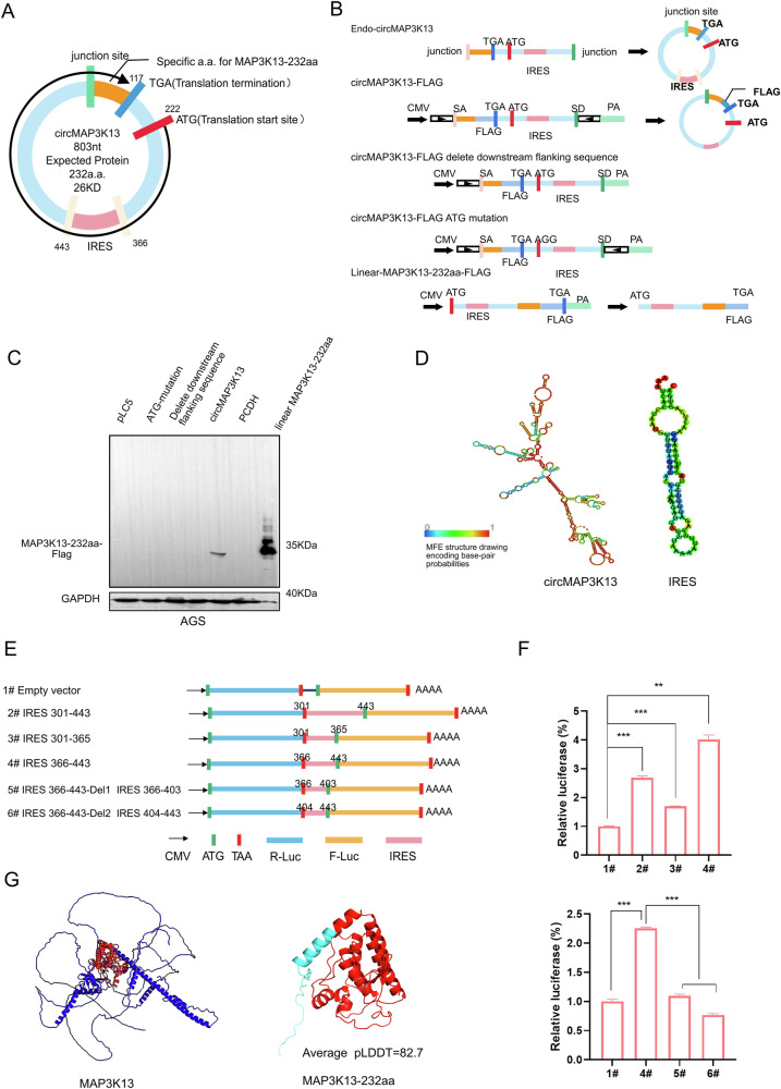
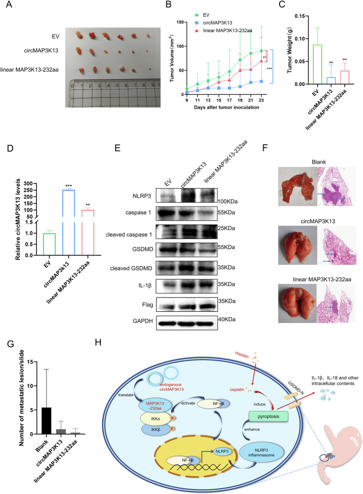
Gastric cancer (GC) is one of the most common and lethal malignancies in developing countries, with particularly high prevalence in China. Circular RNAs (circRNAs) have garnered increasing attention for their roles in disease pathogenesis. While circRNAs can be translated, there have been few investigations into the biological functions of "translatable circRNAs" in the initiation and progression of gastric adenocarcinoma. In this study, we identified a circRNA, circMAP3K13, which inhibits the proliferation and migration of GC cells. CircMAP3K13 was found to encode a previously unreported 26 kDa protein, designated MAP3K13-232aa. Mechanistically, MAP3K13-232aa binds directly to the kinase domain of IKKα and enhances its activity, thereby promoting NF-κB signaling. This activation leads to upregulation of NLRP3 and increased cisplatin-induced pyroptosis in GC cells. Moreover, MAP3K13-232aa enhances pyroptosis and reduces tumorigenicity and metastasis in vivo. Taken together, both circMAP3K13 and its encoded protein MAP3K13-232aa represent potential therapeutic targets in GC.


Conflict of interest statement

Competing interests: The authors declare no competing interests. Ethical approval and consent to participate: The study was approved by the Institutional Review Board of Shenzhen University Health Science Center. Written informed consent was obtained from all patients. All methods in the study were performed in accordance with the guidelines of The First Affiliated Hospital of Shenzhen University.

Figures

Fig. 1
Fig. 1. RNA-seq and Ribo-seq screening of differentially expressed circRNAs in GC.
To construct circRNA expression profiles, RNA-seq and Ribo-seq were performed on two normal human gastric mucosal epithelial cell lines (GES-1 and HFE-145) and four human gastric cancer (GC) cell lines (AGS, MKN-28, SGC-7901, and BGC-823) using the Illumina HiSeq™ 2500 platform. A Classification of circRNAs detected by RNA-seq, grouped by circRNA type. B Distribution of circRNAs counts, as detected by RNA-seq and Ribo-seq. C Length distribution of circRNAs, as identified by RNA-seq and Ribo-seq. D Heatmap showing gene expression profiles from RNA-seq and Ribo-seq analyses across GC and normal cell lines. E KEGG pathway enrichment analysis showing the top 20 significantly enriched pathways in differentially expressed genes between normal (GES-1 and HFE-145) and GC cells. F Volcano plots of differentially expressed genes in GES-1 vs. GC and HFE-145 vs. GC comparisons, highlighting MAPK pathway-associated genes (left and middle). RNA-seq data from five pairs of GC and adjacent normal tissues. circMAP3K13 (right, black arrow) was detected in five paired GC tissues and adjacent normal tissues by RNA-seq.
Fig. 2
Fig. 2. Expression of circMAP3K13 in GC.
A Schematic of the MAP3K13 gene (upper), consisting of 14 exons; circMAP3K13 is encoded by exons 3–7. Divergent and convergent primers were designed to detect circMAP3K13 and linear MAP3K13, respectively. DNA agarose gel detecting circMAP3K13 in AGS cells (lower, left) Sanger sequencing results (lower, middle), and the junction site of circMAP3K13 between exons 3 and 7 (lower, right). B Total RNA was treated with RNase R, and the digested RNA was analyzed by qPCR to assess the expression of circMAP3K13 and MAP3K13. DNA agarose gel displays the qPCR products. C FISH was performed to determine the localization of circMAP3K13 (red) in AGS and MKN-45 cells. Co-staining with the endoplasmic reticulum marker Calnexin (green) was carried out in MKN-45 cells. Nuclei are counterstained with DAPI (blue). D qPCR analysis of circMAP3K13 in nuclear and cytoplasmic fractions of AGS cells. 18S and U6 served as cytoplasmic and nuclear controls, respectively. E Bar chart showing qPCR results for circMAP3K13 expression in 65 paired GC (pink) and adjacent non-tumor tissues (gray); P = 0.002. F CircMAP3K13 expression in GC cell lines. G CircMAP3K13 expression in GC patients. The percentage of patients with low expression in stage I gastric cancer was higher than that of patients with low expression in stages Ⅱ, Ⅲ and Ⅳ. The Chi-square test was used to analyze the data. H circMAP3K13 expression in tumors with different infiltration degrees (n = 48 patients). The higher the expression of circMAP3K13, the deeper the invasion depth of the tumor. T1: invasion into the mucosal or muscular layer; T2: invasion into the subserosal layer or outer serosa; T3: penetration of the gastric wall with serosal involvement; T4: invasion beyond the serosa or into adjacent structures. I Association between circMAP3K13 expression and lymph node metastasis. N0: no metastasis; N1: 1–2 nodes; N2: 3–6 nodes; N3: 7 or more nodes. Chi-square test was used. J Receiver operating characteristic curve for circMAP3K13 expression in GC versus adjacent tissues. The reference line (red dotted), ROC curve (green), and cutoff point (red dot) are shown. If not otherwise specified, data were analyzed using two-tailed Student’s t-test. *P < 0.05; **P < 0.01; ***P < 0.001.
Fig. 3
Fig. 3. circMAP3K13 harbors an active IRES sequence, and can be translated into a novel protein.
A Schematic of circMAP3K13, which consists of 803 nucleotides and is predicted to encode a novel protein of approximately 26 kDa. B Schematic representation of plasmid constructs. The circMAP3K13-FLAG plasmid overexpresses circMAP3K13 with a 3×FLAG tag. The downstream flanking sequence deletion mutant lacks the downstream complementary sequence required for circularization. The ATG-mutation construct disrupts the start codon, altering the open reading frame. The linear-MAP3K13-232aa-FLAG plasmid expresses the same ORF of circMAP3K13 in a linear form. C Western blot analysis of circMAP3K13-FLAG and linear-MAP3K13-232aa-FLAG. D RNAfold software prediction of the secondary structure of circMAP3K13 and a putative internal ribosome entry site (IRES). E Schematic of five recombinant plasmids generated to assess IRES activity by dual-luciferase reporter assay. F Relative luciferase activity of plasmids 1–6. G AlphaFold3–based 3D structure prediction of MAP3K13-232aa. The MAP3K13-232aa sequence is highlighted in red within the full MAP3K13 protein (left) and the specific MAP3K13-232aa peptide is shown in cyan (right). Unless otherwise specified, statistical analysis was performed using a two-tailed Student’s t-test. **P < 0.01; ***P < 0.001.
Fig. 4
Fig. 4. CircMAP3K13 encodes a novel protein, MAP3K13-232aa.
A Mass spectrometry (IP-MS) following immunoprecipitation was conducted to identify the unique peptide of MAP3K13-232aa. B A commercial MAP3K13 antibody for MAP3K13 was used to detect the overexpressed MAP3K13-232aa and endogenous MAP3K13-232aa by immunoblotting. C MAP3K13-232aa expression level was analyzed in nine pairs of GC and paired adjacent normal tissues by western blot. N normal tissue, T tumor tissue. D Immunoblot analysis of MAP3K13-232aa across various cell lines, including GC cell lines (NCI-N87, MKN-45, HGC-27, AGS, MKN-28), a normal gastric epithelial cell line (GES-1), and 293 T cells. GAPDH served as the loading control. E Schematic of three siRNAs targeting circMAP3K13. F qPCR results of circMAP3K13 expression after transfection with si-1, si-2, and si-3. G Western blot analysis of MAP3K13-232aa protein as well as full-length MAP3K13 after circMAP3K13 siRNA transfection. GAPDH served as the loading control. Unless otherwise specified, statistical significance was assessed using a two-tailed Student’s t-test. *P < 0.05; **P < 0.01; ***P < 0.001; ****P < 0.0001.
Fig. 5
Fig. 5. circMAP3K13 suppresses the proliferation and migration of GC cells.
A Cell proliferation was determined by using EdU staining (red) after overexpression of MAP3K13-232aa in AGS cells and B after silencing circMAP3K13 with siRNAs transfection in HGC-27 cells. The nuclei are counterstained with DAPI (blue). Scale bar = 10 μm. C Quantification of EdU-positive cells from (A, B). D Wound healing assay was performed after circMAP3K13 and MAP3K13-232aa overexpression. Scale bar = 100 μm. E Wound healing assay was performed after knockdown of circMAP3K13 in HGC-27 cells. Scale bar = 200 μm. F Quantification of wound healing assays is shown. G Transwell assay was conducted after overexpressing MAP3K13-232aa in AGS cells. Scale bar = 100 μm. H Transwell assay was conducted after transfection of circMAP3K13-targeting siRNAs in HGC-27 cells. Scale bar = 100 μm. I Quantification of Transwell invasion assay results is shown. Unless otherwise specified, statistical significance was assessed using a two-tailed Student’s t-test. *P < 0.05; **P < 0.01; ***P < 0.001; ****P < 0.0001.
Fig. 6
Fig. 6. MAP3K13-232 directly binds to the kinase domain of IKKα.
A Proteins interacting with MAP3K13-232aa were identified by immunoprecipitation followed by mass spectrometry (IP-MS). A total of 2305 proteins were pulled down by anti-flag antibody from lysates of cells transfected with circMAP3K13 or empty vector using an anti-FLAG antibody. Of these, 435 proteins were unique to the circMAP3K13 group. Differentially abundant proteins (fold change >2) were subjected to KEGG pathway enrichment analysis. B Fifty-three proteins with a fold change >13, including the IKKα, with fold changes >13 were enriched in cancer-related pathways; several enriched pathways identified in the enrichment analysis are shown. C The interaction between MAP3K13-232aa and IKKα was modeled using Alphafold3. IKKα is shown in green, and the purple structure is MAP3K13-232aa. Chemical interactions were visualized using PyMOL. Yellow dotted lines represent hydrogen bonds, red dotted lines indicate hydrophobic interactions, and blue dotted lines denote salt bridges. D Co-immunoprecipitation experiments were performed to detect the interaction between MAP3K13-232aa and IKKα. E Schematic of the five GST-tagged IKKα recombinant protein constructs used in the GST pull-down assay: GST-WT (full-length recombinant GST-IKKα); GST-IKKα-ΔNEMO (lacking the NEMO-binding region); GST-IKKα-ΔKinase (kinase domain of GST-IKKα was deleted); GST- IKKα-Kinase (only the kinase domain); GST-IKKα-NEMO (only included the NEMO-binding region of GST-IKKα and lacked the kinase domain, lacking the kinase and leucine zipper regions). F Five “GST-tagged constructs and a His-tagged MAP3K13-232aa construct were expressed in E. coli BL21(DE3). Purified proteins were incubated to form immunocomplexes, followed by western blotting using anti-GST and anti-His antibodies. G AGS cells were transfected with ATG-mutation, circMAP3K13, wild-type circMAP3K13, empty vector (PCDH), linear MAP3K13-232aa, or kinase-dead MAP3K13-232aa. Whole-cell lysates were collected and subjected to immunoprecipitation for kinases enrichment. In vitro kinase assay was performed. Upper panel: IP FLAG; middle and lower panels: IP IKKα. Substrates used were IκB for IKKα and MBP for MAP3K13-232aa. Unless otherwise specified, statistical analysis was performed using a two-tailed Student’s t-test. *P < 0.05; **P < 0.01; ***P < 0.001.
Fig. 7
Fig. 7. MAP3K13-232aa enhances cisplatin-induced pyroptosis through activating the NF-κB signaling pathway.
A Western blot analysis of AGS cells transfected with circMAP3K13 overexpression plasmids. Cells were treated with 20 ng/mL TNFα for 5 min prior to protein collection. NF-κB signaling pathway-related proteins were detected by western blot. B Detection of NF-κB downstream target proteins in AGS cells transfected with circMAP3K13 overexpression plasmids. C AGS cells transfected with either an empty vector or circMAP3K13 overexpression plasmid circMAP3K13 groups were treated with LPS (2 μg/mL) for 4 h and nigericin (20 μM) for 1 h. Red arrows indicate protrusive vesicles characteristic of pyroptosis. D LDH levels in the culture supernatant were measured in AGS cells under the same treatment conditions to evaluate the extent of pyroptosis. E NLRP3 mRNA expression was assessed in AGS cells after induction of pyroptosis. F Western blot analysis was used to detect NLRP3 protein expression following pyroptosis induction after circMAP3K13 overexpression and knockdown. Western blot analysis of pyroptosis-related proteins in MKN-45 (G) or N87 (H) cells, which were treated with cisplatin (1 μg/mL) for 48 h. I Rescue assay showing the effect of MAP3K13-232aa overexpression following circMAP3K13 knockdown on pyroptosis-associated protein expression. Unless otherwise specified, statistical analysis was performed using a two-tailed Student’s t-test. *P < 0.05; **P < 0.01; ***P < 0.001; ****P < 0.0001.
Fig. 8
Fig. 8. Overexpression of circMAP3K13 overexpression curbs tumor growth in vivo.
A Tumorigenic models were established by subcutaneous injection of nude mice with 1 × 10⁶ MKN-45 cells which were stably transfected with circMAP3K13, MAP3K13-232aa, or empty vector. B, C Tumor volume was measured at regular intervals in each group. C Tumor weight was assessed at the endpoint. D Quantitative PCR and western blot analyses were performed on tumor tissues to determine the expression levels of circMAP3K13 and MAP3K13-232aa. E Pyroptosis-related proteins were detected in tumor lysates to evaluate pyroptotic activity. F Hematoxylin and eosin staining to identify metastases in the lungs of nude mice. G Quantification of metastatic lung nodules in each group. H Schematic model of the proposed mechanism: circMAP3K13 encodes MAP3K13-232aa, which binds to the kinase domain of IKKα to activate the NF-κB signaling and promote cisplatin-induced pyroptosis. Unless otherwise specified, statistical analysis was performed using a two-tailed Student’s t-test. **P < 0.01; ***P < 0.001.

Similar articles

References

    1. Johnston FM, Beckman M. Updates on management of gastric cancer. Curr Oncol Rep. 2019;21:67. - PubMed
    1. Matsuoka T, Yashiro M. Biomarkers of gastric cancer: current topics and future perspective. World J Gastroenterol. 2018;24:2818–32. - PMC - PubMed
    1. Sanger HL, Klotz G, Riesner D, Gross HJ, Kleinschmidt AK. Viroids are single-stranded covalently closed circular RNA molecules existing as highly base-paired rod-like structures. Proc Natl Acad Sci USA. 1976;73:3852–6. - PMC - PubMed
    1. Hsu MT, Cocaprados M. Electron-microscopic evidence for the circular form of RNA in the cytoplasm of eukaryotic cells. Nature. 1979;280:339–40. - PubMed
    1. Altesha MA, Ni T, Khan A, Liu K, Zheng X. Circular RNA in cardiovascular disease. J Cell Physiol. 2019;234:5588–5600. - PubMed

MeSH terms