

Subjective cognitive decline in major depressive patients is associated with altered entropy and connectivity changes of temporal and insular region
- PMID: 40890120
- PMCID: PMC12402481
- DOI: 10.1038/s41398-025-03518-w
Subjective cognitive decline in major depressive patients is associated with altered entropy and connectivity changes of temporal and insular region
Abstract
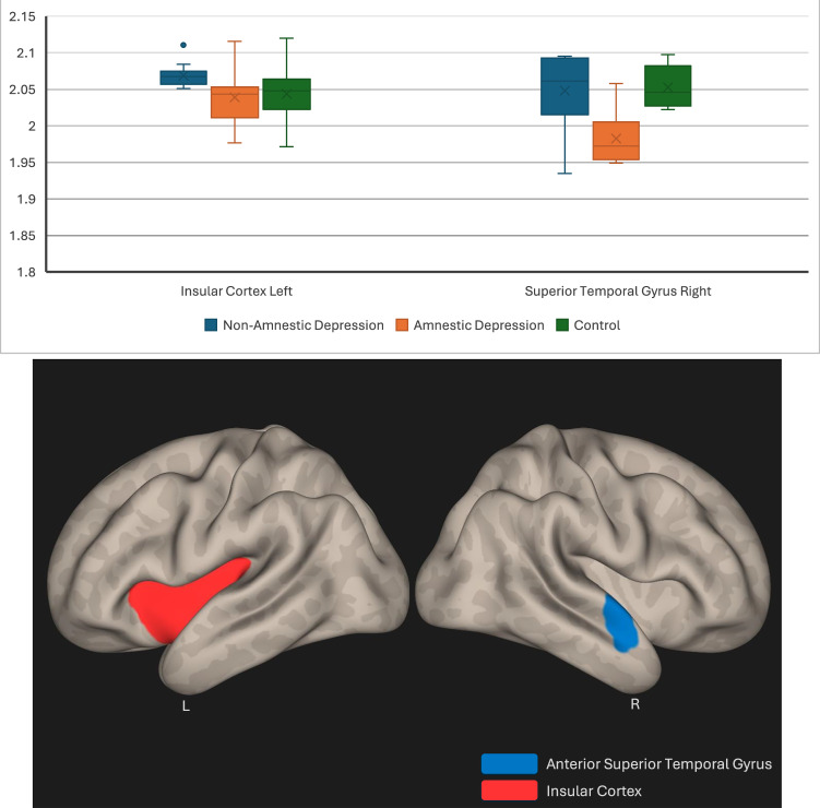
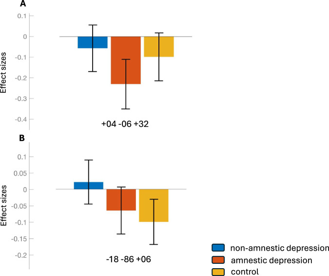
Depressive cognitive impairment is seen in a significant number of patients with depression. However, it remains challenging to differentiate between patients with amnestic (those with subjective cognitive impairment complaints) and non-amnestic major depressive disorder, highlighting the urgent need for additional objective tools to help classify these patients more accurately. We analyzed cognitive state, alterations in regional entropy and functional connectivity measures of the brain between patients with major depression and healthy controls. The depressed cohort was categorized as either "amnestic" or "non-amnestic," depending on self-reported experiences of forgetfulness. The superior temporal region and insula exhibited altered entropy and connectivity measures in individuals with depression and subjective cognitive impairment, which was correlated with impaired executive functions, a pattern not being evident in the control group. Our findings support the notion that insular and superior temporal entropic alterations are linked to subjective cognitive changes in the pathology of depression. These regions also hold potential as biomarkers for determining the underlying objective cognitive deficits in subjective cognitive complaints in patients with major depressive disorder (MDD). This underscores the need for improved diagnostic approaches and the implementation of practical dynamic neuroimaging modalities capable of addressing the current challenges in diagnosing subjective cognitive impairment in MDD, offering promise for the future management of patients with depression.
© 2025. The Author(s).
Conflict of interest statement
Competing interests: The authors declare no competing interests. Ethics approval and consent to participate: The study protocol was approved by the Ethics Committee of Istanbul Medipol University and written informed consent forms were obtained from participants after thoroughly informing them about the study protocol (Ethical Number: 10840098‐604.01.01‐E.19402). All methods were carried out in accordance with relevant guidelines and regulations. Consent for publication: All participants signed consent forms and agreed to participate in this research.
Figures

References
-
- Austin MP, Mitchell P, Goodwin GM. Cognitive deficits in depression: possible implications for functional neuropathology. Br J Psychiatry. 2001;178:200–6. - PubMed
-
- Bennett S, Thomas AJ. Depression and dementia: cause, consequence or coincidence? Maturitas. 2014;79:184–90. - PubMed
-
- Rubin R. Exploring the relationship between depression and dementia. JAMA. 2018;320:961. - PubMed
-
- Gao Y, Huang C, Zhao K, Ma L, Qiu X, Zhang L, et al. Retracted: Depression as a risk factor for dementia and mild cognitive impairment: a meta‐analysis of longitudinal studies. Int J Geriatr Psychiatry. 2013;28:441–9. - PubMed
MeSH terms
LinkOut - more resources
Full Text Sources
