

Computer vision based efficient segmentation and classification of multi brain tumor using computed tomography images
- PMID: 40890232
- PMCID: PMC12402189
- DOI: 10.1038/s41598-025-16825-5
Computer vision based efficient segmentation and classification of multi brain tumor using computed tomography images
Abstract
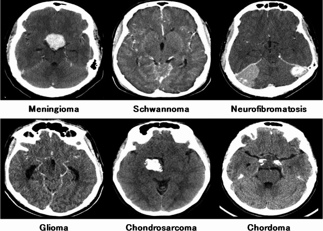
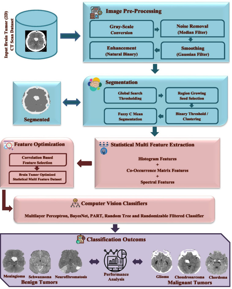
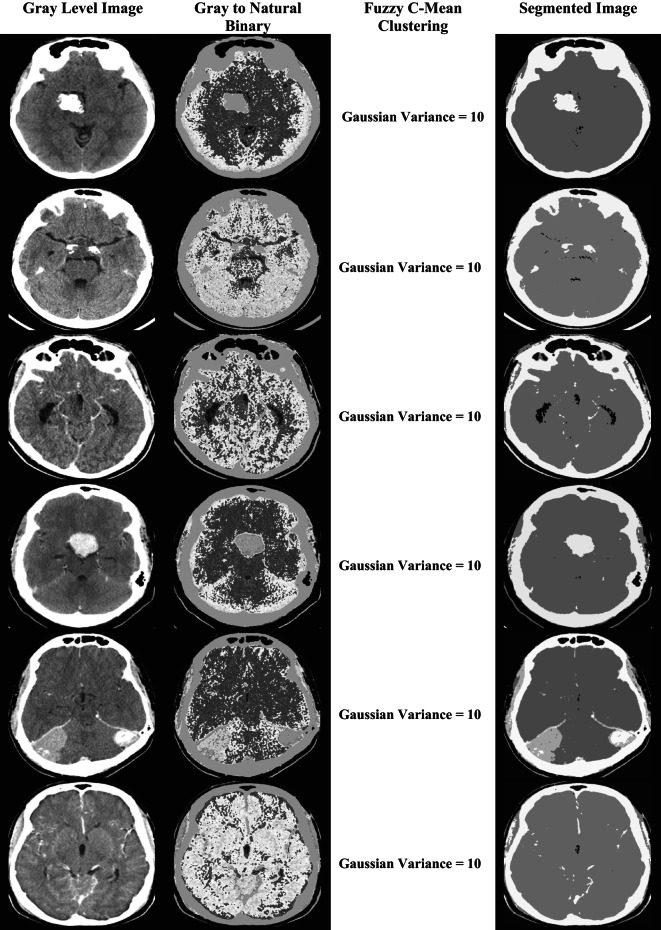
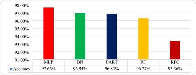
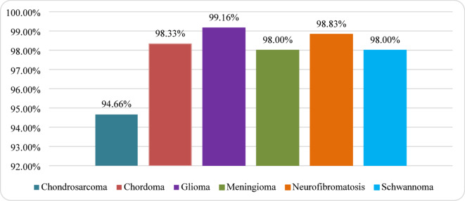
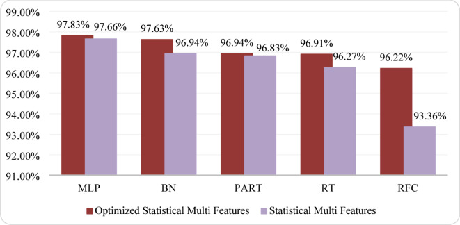
This study aims to highlight the effectiveness of computer vision (CV) techniques in classifying brain tumors using a comprehensive dataset consisting of computed tomography (CT) scans. The proposed framework comprises six types of brain tumors, including benign tumors (Meningioma, Schwannoma, and Neurofibromatosis) and malignant tumors (Glioma, Chondrosarcoma, and Chordoma). The acquired images underwent pre-processing steps to enhance the dataset's quality, including noise reduction through median and Gaussian filters and region of interest (ROIs) extraction using an automated binary threshold-based fuzzy c-means segmentation (ABTFCS) approach. A total of 900 CT-scan images were utilized, 150 images per tumor class, each with a size of 512 × 512 pixels, and 4 ROIs taken per image, so the total dataset size is 3600 (900 × 4) attributes. After pre-processing, the dataset was further analysed to extract 135 statistical multi-features for each ROI. An optimized set of 12 statistical multi-features was selected to identify the most relevant features using a feature selection technique based on correlation. For the classification stage, the optimized statistical multi-feature dataset was evaluated using five computer vision classifiers: multilayer perceptron (MLP), BayesNet, PART, random tree, and randomizable filtered classifier, employing a 10-fold cross-validation method. Among these classifiers, MLP with fine-tuned hyperparameters achieved a promising accuracy rate of 97.83%.
Keywords: ABTFCS; Brain tumor; Computer vision; Multilayer perceptron; Optimized statistical multi-features.
© 2025. The Author(s).
Conflict of interest statement
Declarations. Competing interests: The authors declare no competing interests.
Figures


References
-
- Tiwari, S. & Talreja, S. Do you think disease and disorder are same? –Here is the comparative review to brash up your knowledge. J. Pharm. Sci. Res.12 (4), 462–468 (2020).
-
- Vilella, F. & Simon, C. Reproductive medicine, as seen through single-cell glasses. Fertil. Steril.115 (2), 296–297 (2021). - PubMed
MeSH terms
LinkOut - more resources
Full Text Sources
Medical
Research Materials
