

CT-based deep learning radiomics model for predicting proliferative hepatocellular carcinoma: application in transarterial chemoembolization and radiofrequency ablation
- PMID: 40890681
- PMCID: PMC12403943
- DOI: 10.1186/s12880-025-01913-9
CT-based deep learning radiomics model for predicting proliferative hepatocellular carcinoma: application in transarterial chemoembolization and radiofrequency ablation
Abstract
Objectives: Proliferative hepatocellular carcinoma (HCC) is an aggressive tumor with varying prognosis depending on the different disease stages and subsequent treatment. This study aims to develop and validate a deep learning radiomics (DLR) model based on contrast-enhanced CT to predict proliferative HCC and to implement risk prediction in patients treated with transarterial chemoembolization (TACE) and radiofrequency ablation (RFA).
Materials and methods: 312 patients (mean age, 58 years ± 10 [SD]; 261 men and 51 women) with HCC undergoing surgery at two medical centers were included, who were divided into a training set (n = 182), an internal test set (n = 46) and an external test set (n = 84). DLR features were extracted from preoperative contrast-enhanced CT images. Multiple machine learning algorithms were used to develop and validate proliferative HCC prediction models in training and test sets. Subsequently, patients from two independent new sets (RFA and TACE sets) were divided into high- and low-risk groups using the DLR score generated by the optimal model. The risk prediction value of DLR scores in recurrence-free survival (RFS) and time to progression (TTP) was examined separately in RFA and TACE sets.
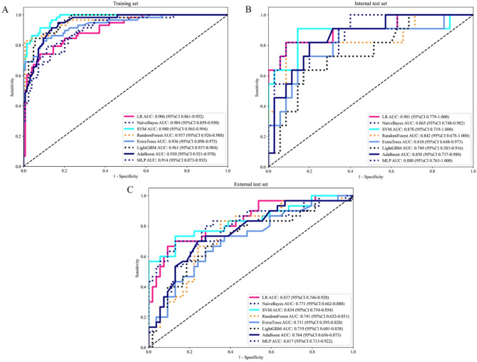
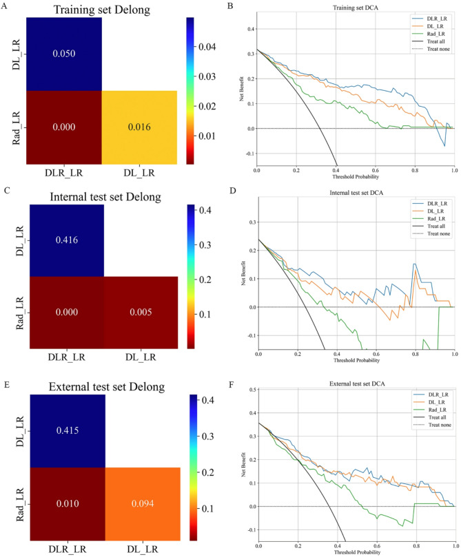
Results: The DLR proliferative HCC prediction model demonstrated excellent predictive performance with an AUC of 0.906 (95% CI 0.861–0.952) in the training set, 0.901 (95% CI 0.779–1.000) in the internal test set and 0.837 (95% CI 0.746–0.928) in the external test set. The DLR score effectively enables risk prediction for patients in RFA and TACE sets. For the RFA set, the low-risk group had significantly longer RFS compared to the high-risk group (P = 0.037). Similarly, the low-risk group showed a longer TTP than the high-risk group for the TACE set (P = 0.034).
Conclusions: The DLR-based contrast-enhanced CT model enables non-invasive prediction of proliferative HCC. Furthermore, the DLR risk prediction helps identify high-risk patients undergoing RFA or TACE, providing prognostic insights for personalized management.
Supplementary Information: The online version contains supplementary material available at 10.1186/s12880-025-01913-9.
Keywords: Contrast-enhanced CT; Deep learning radiomics; Hepatocellular carcinoma; Radiofrequency ablation (RFA); Transarterial chemoembolization (TACE).
Conflict of interest statement
Declarations. Ethics approval and consent to participate: This study was performed in line with the principles of the Declaration of Helsinki. Approval was granted by the Ethics Committee of Tianjin First Central Hospital. Written informed consent was waived from each patient due to the retrospective study. Competing interests: The authors declare no competing interests.
Figures


References
-
- Calderaro J, Ziol M, Paradis V, Zucman-Rossi J. Molecular and histological correlations in liver cancer. J Hepatol. 2019;71:616–30. - PubMed
Grants and funding
LinkOut - more resources
Full Text Sources
Miscellaneous
