. 2025 Sep 1;32(1):82.
doi: 10.1186/s12929-025-01164-4.

Synaptic accumulation of GluN2B-containing NMDA receptors mediates the effects of BDNF-TrkB signalling on synaptic plasticity and in hyperexcitability during status epilepticus

Affiliations

Synaptic accumulation of GluN2B-containing NMDA receptors mediates the effects of BDNF-TrkB signalling on synaptic plasticity and in hyperexcitability during status epilepticus

Pasqualino De Luca et al. J Biomed Sci. .

Abstract

Background: Brain-derived neurotrophic factor (BDNF) is a key mediator of synaptic plasticity and memory formation in the hippocampus. However, the BDNF-induced alterations in the glutamate receptors coupled to the plasticity of glutamatergic synapses in the hippocampus have not been elucidated. In this work we investigated the putative role of GluN2B-containing NMDA receptors in the plasticity of glutamatergic synapses induced by BDNF.

Methods: The effects of BDNF on the surface expression of GluN2B-containing NMDA receptors was investigated in cultured hippocampal neurons and in hippocampal synaptoneurosomes by immunocytochemistry under non-permeabilizing conditions, using an antibody that binds to an extracellular epitope. Long term potentiation of hippocampal CA1 synapses was induced by using θ-burst stimulation. Epileptic seizures were induced using the Li+-pilocarpine model of temporal lobe epilepsy. Pyk2 phosphorylation was assessed by western blot with a phosphospecific antibody.

Results: Stimulation of hippocampal synaptoneurosomes with BDNF led to a significant time-dependent increase in the synaptic surface expression of GluN2B-containing NMDA receptors as determined by immunocytochemistry with colocalization with pre- (vesicular glutamate transporter) and post-synaptic markers (PSD-95). Similarly, BDNF induced the synaptic accumulation of GluN2B-containing NMDA receptors at the synapse in cultured hippocampal neurons, by a mechanism sensitive to the PKC inhibitor GӦ6983. The effects of PKC may be mediated by phosphorylation of Pyk2, as suggested by western blot experiments analyzing the phosphorylation of the kinase on Tyrosine 402. GluN2B-containing NMDA receptors mediated the effects of BDNF in the facilitation of the early phase of long-term potentiation (LTP) of hippocampal CA1 synapses induced by θ-burst stimulation, since the effect of the neurotrophin was abrogated in the presence of the GluN2B inhibitor Co 101244. In the absence of BDNF, the GluN2B inhibitor did not affect LTP. Surface accumulation of GluN2B-containing NMDA receptors was also observed in hippocampal synaptoneurosomes isolated from rats subjected to the pilocarpine model of temporal lobe epilepsy, after reaching Status Epilepticus, an effect that was inhibited by administration of the TrkB receptor inhibitor ANA-12.

Conclusion: Together, these results show that the synaptic accumulation of GluN2B-containing NMDA receptors mediate the effects of BDNF in the plasticity of glutamatergic synapses in the hippocampus.

Keywords: BDNF; Epilepsy; Hippocampal synapses; LTP; NMDA receptors.


Conflict of interest statement

Declarations. Ethics approval and consent to participate: Not applicable. Consent for publication: Not applicable. Competing interests: The authors declare that they have no competing interests.

Figures
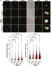
Fig. 1
Fig. 1
BDNF-induced increase in the expression of GluN2B-containing NMDAR in hippocampal synaptoneurosomes. A Representative images of hippocampal synaptoneurosomes (prepared from adult Sprague–Dawley rats 6–8 weeks old) incubated with BDNF (50 ng/mL). Synaptoneurosomes were immunoassayed for GluN2B using an antibody against an extracellular epitope in the GluN2B N-terminus and immunoassayed for vGluT1and PSD-95. Merge scale bar, 10 μm. Insert scale bar, 0.5 µm. Images illustrated in A were analyzed for the GluN2B integrated density (B) and GluN2B mean gray value (C). Data are the means ± SEM of 779—840 synaptoneurosomes per condition, in at least three independent experiments performed in different preparations. ****p < 0.0001, by Kruskal–Wallis’s test and Dunn’s multiple comparisons test
Fig. 2
Fig. 2
Stimulation of hippocampal neurons with BDNF for 30 min increases the synaptic expression of GluN2B-containing NMDAR. A Representative images of hippocampal neurons (DIV 14-15) that were stimulated with BDNF (50 ng/ml for 10 or 30 min), as indicated. Neurons were then live-immunoassayed for GluN2B using an antibody against an extracellular epitope in the GluN2B N-terminus, fixed, and then further immunoassayed for PSD-95, vGluT1, and MAP2. Scale bar, 5 μm. Images illustrated in (A), were analyzed for the total number (B), intensity (C), and area (D) of surface GluN2B puncta per µm. Synaptic (PSD-95- and vGluT1-colocalized) surface GluN2B number (E), area (F), and intensity (G) of puncta per density of excitatory synapses (number of puncta PSD-95–vGluT1 colocalized per µm), were also analyzed. Data are normalized to the means of the control and are the means ± SEM of 43–45 cells per condition, from at least three independent experiments performed in different preparations. *p < 0.05, **p < 0.01 by one-way analysis of variance (ANOVA) followed by Bonferroni post-test
Fig. 3
Fig. 3
BDNF-induced activation/phosphorylation of Pyk2 is dependent on PKC activation. Representative western blots and analysis (A, E). Hippocampal neurons (14-15 DIV) were stimulated or not with BDNF (50 ng/mL) during 10- and 30-min (B, C, D) and with or without GÖ6983 where indicated (F, G, H). Proteins were extracted then resolved and analyzed by immunoblot, using antibodies against pPyk2, Pyk2, and β-Tubulin. In this analysis, the pPyk2 levels were normalized to β-Tubulin (C) and total Pyk2 (B). Data are means ± SEM of at least three different experiments, performed in independent preparations. For each preparation, the data were normalized to the value of the control condition and expressed as a percentage of the control. *p < 0.05, **p < 0.01, ***p < 0.001 by one-way analysis of variance (ANOVA) followed by Bonferroni post-test
Fig. 4
Fig. 4
BDNF-induced increase in the synaptic expression of GluN2B-containing NMDAR is sensitive to PKC inhibition. A Representative images of hippocampal neurons (DIV 14-15) pre-incubated with GÖ 6983 (100 nM) or vehicle (DMSO; 1:1000 dilution for 40 min) and then either maintained under the same conditions or stimulated with BDNF (50 ng/ml for 30 min), as indicated. Neurons were live-immunoassayed for GluN2B using an antibody against an extracellular epitope in the GluN2B N-terminus, fixed, and then further immunoassayed for PSD-95, vGluT1, and MAP2. Scale bar, 5 μm. Images illustrated in A were analyzed for the total number (B), area (C), and intensity (D) of surface GluN2B puncta per µm. Synaptic (PSD-95- and vGluT1-colocalized) surface GluN2B number (E), area (F), and intensity (G) of puncta per µm of excitatory synapses (number of puncta PSD-95–vGluT1 colocalized per µm), were also analyzed. Data are normalized to the mean of the DMSO control and are the means ± SEM of 28-30 cells per condition, in at least three independent experiments performed in different preparations. ***p < 0.001, ****p < 0.0001 by one-way analysis of variance (ANOVA) followed by Bonferroni post-test
Fig. 5
Fig. 5
BDNF-induced enhancement of LTP is antagonized by GluN2B-containing NMDAR inhibition. A Averaged time course changes in fEPSP slope induced by a θ-burst stimulation (% of change) in the absence (black dots) or in the presence of 20 ng/ml BDNF (red dots) (A, C n = 7; G, I n = 6) or Co 101244 alone (D, F n = 5). Note that in G-I BDNF was tested in the presence of Co 101244. The ordinates represent normalized fEPSP slopes, where the averaged slopes recorded for 10 min before burst stimulation were set to 0%, and the abscissa represents the recording time. Representative traces (B, E, and H). Each trace is the average of consecutive responses obtained at the indicated periods in the time-course panel on the left, i.e. before (1 and 3) or 60 min after (2 and 4) LTP induction, and is composed of the stimulus artifact, followed by the presynaptic volley and the fEPSP. Traces 1 and 2 were obtained in the absence of BDNF and traces 3 and 4 in its presence from a second pathway in the same slice. Traces recorded from the same pathway before and after LTP induction are superimposed. C, F, I Bar graphs show the magnitude of LTP (change in the fEPSP slope over time) induced by θ-burst stimulation in relation to pre-burst values (0%) in the same hippocampal slices. Statistical analysis for the LTP magnitude was performed by the t-test. (*p < 0.05)
Fig. 6
Fig. 6
The increase in the expression of GluN2B-containing NMDAR in hippocampal synaptoneurosomes during epileptogenesis requires TrkB signaling. A Experimental design for the lithium-pilocarpine model of Status Epilepticus. B Representative images of hippocampal synaptoneurosomes (prepared from adult Sprague–Dawley rats 6–8 weeks old, treated with saline, saline and ANA-12, Pilocarpine or Pilocarpine and ANA-12). Synaptoneurosomes were live-immunoassayed for GluN2B using an antibody against an extracellular epitope in the GluN2B N-terminus and immunoassayed for vGluT1 and PSD-95. Merge scale bar, 10 μm. Insert scale bar, 0.5 µm. Images illustrated in (B) were analyzed for the GluN2B integrated density (C) and GluN2B mean gray value (D). Data are the means ± SEM of 779-840 synaptoneurosomes per condition, from at least four animals for each experimental condition. ****p < 0.0001, **p < 0.01 as determined by Kruskal Wallis’s test and Dunn’s multiple comparisons test. Representative western blot and analysis (E–G). Proteins were extracted from synaptoneurosomes prepared from the same animals used in the immunocytochemistry experiments. For the immunoblot, antibodies against pTrkB, total TrKB and β-Tubulin were used. In this analysis, pTrkB levels were normalized to total TrkB levels and total TrkB levels were normalized to β-Tubulin. Data are means ± SEM of at least four animals for each experimental condition. *p < 0.05, by one-way analysis of variance (ANOVA) followed by Dunnett’s post-test

Similar articles

References

    1. Afonso P, De Luca P, Carvalho RS, Cortes L, Pinheiro P, Oliveiros B, Almeida RD, Mele M, Duarte CB. BDNF increases synaptic NMDA receptor abundance by enhancing the local translation of Pyk2 in cultured hippocampal neurons. Sci Signal. 2019;12(586):eaav3577. - PubMed
    1. Akashi K, Kakizaki T, Kamiya H, Fukaya M, Yamasaki M, Abe M, Natsume R, Watanabe M, Sakimura K. NMDA receptor GluN2B (GluR epsilon 2/NR2B) subunit is crucial for channel function, postsynaptic macromolecular organization, and actin cytoskeleton at hippocampal CA3 synapses. J Neurosci. 2009;29(35):10869–82. - PMC - PubMed
    1. Andersson O, Stenqvist A, Attersand A, von Euler G. Nucleotide sequence, genomic organization, and chromosomal localization of genes encoding the human NMDA receptor subunits NR3A and NR3B. Genomics. 2001;78(3):178–84. - PubMed
    1. Baez MV, Cercato MC, Jerusalinsky DA. NMDA receptor subunits change after synaptic plasticity induction and learning and memory acquisition. Neural Plast. 2018:5093048. - PMC - PubMed
    1. Barria A, Malinow R. Subunit-specific NMDA receptor trafficking to synapses. Neuron. 2002;35(2):345–53. - PubMed

MeSH terms

LinkOut - more resources