. 2025 Nov 3;222(11):e20242403.
doi: 10.1084/jem.20242403. Epub 2025 Sep 2.

Discovery of potent and selective inhibitors of human NLRP3 with a novel mechanism of action

Affiliations

Discovery of potent and selective inhibitors of human NLRP3 with a novel mechanism of action

Kevin Wilhelmsen et al. J Exp Med. .

Abstract

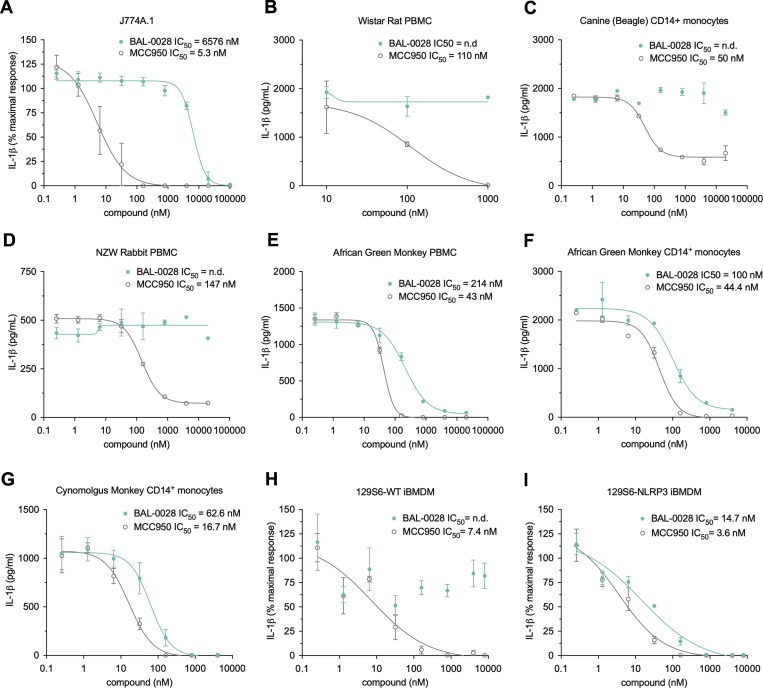
The NLRP3 inflammasome is an intracellular protein complex that causes inflammation via the release of IL-1β and pyroptosis. NLRP3 activation is associated with many age-related inflammatory diseases, and NLRP3 inhibition is a promising therapeutic strategy. We previously performed a DNA-encoded library screen to identify novel NLRP3-binding molecules. Herein we describe the characterization of BAL-0028 as a potent and specific inhibitor of NLRP3 signaling. Notably, BAL-0028 is a poor inhibitor of mouse NLRP3 but inhibits human and primate NLRP3 with nanomolar potency. Using cellular and biochemical analyses, we demonstrate that BAL-0028 binds to the NLRP3 NACHT domain at a site that is distinct from the MCC950-binding pocket. Using humanized NLRP3 mice, we show that a derivative of BAL-0028, BAL-0598, inhibits NLRP3 activation in vivo in a peritonitis model. Finally, we demonstrate that both BAL-0028 and BAL-0598 inhibit select hyperactive NLRP3 mutations associated with autoinflammatory diseases more potently than MCC950. BAL-0028 and BAL-0598 thus represent a new modality for NLRP3 inhibition in inflammatory diseases.


Conflict of interest statement

Disclosures: K. Wilhelmsen, A. Deshpande, S. Tronnes, M. Banicki, M. Cochran, S. Cowdin, R. Montgomery, C.P. Portillo, P. Rubin, and Y. Wang reported grants and nonfinancial support from BioAge Labs, Inc. during the conduct of the study; and personal fees from BioAge Labs, Inc. outside the submitted work. M. Mahanta and T. Salazar reported personal fees and nonfinancial support from BioAge Labs, Inc. during the conduct of the study; and personal fees from BioAge Labs, Inc. outside the submitted work. K. Fortney and G. Hartman reported grants from BioAge Labs, Inc. during the conduct of the study; and personal fees from BioAge Labs, Inc. outside the submitted work. R.E. Hughes reported grants, personal fees, and nonfinancial support from BioAge Labs, Inc. outside the submitted work. M. Geyer reported grants and personal fees from BioAge Labs during the conduct of the study. R.C. Coll reported grants and personal fees from BioAge Labs during the conduct of the study; and personal fees from Viva in vitro diagnostics outside the submitted work. A.N.R. Weber is in the process of applying for a grant on NLRP3 research, but this has not influenced his contribution to this publication. In addition, K. Wilhelmsen, S. Tronnes, M. Mahanta, M. Banicki, M. Cochran, S. Cowdin, K. Fortney, R.E. Hughes, R. Montgomery, C.P. Portillo, P. Rubin, T. Salazar, and Y. Wang had patent numbers 11,702,391 and 11,708,334 issued (BioAge Labs, Inc.); B.F. Py reported a patent to PCT/EP2024/074525 issued; M. Geyer had a patent to 19/089,839 pending; and R.C. Coll had a patent to US 10,538,48 licensed, a patent to PCT/IB2017/053059 licensed, a patent to PCT/EP2017/053498 licensed, and a patent to US 18/906,535 pending. K. Wilhelmsen, A. Deshpande, S. Tronnes, M. Mahanta, M. Banicki, M. Cochran, S. Cowdin, K. Fortney, G. Hartman, R.E. Hughes, R. Montgomery, C.P. Portillo, P. Rubin, T. Salazar, and Y. Wang are employed by BioAge Labs, Inc., a biotechnology company developing NLRP3 inhibitors. M. Geyer and R.C. Coll are consultants for BioAge Labs, Inc. No other disclosures were reported.

Figures

Figure 1.
Figure 1.
BAL-0028 is a potent inhibitor of NLRP3 signaling in multiple human cell types. (A) Structure of BAL-0028. (B–D) Comparison of BAL-0028 and MCC950 in IL-1β release assays from PMA-differentiated THP-1 cells stimulated with LPS and (B) nigericin, (C) ATP, or (D) MSU. (E–I) Comparison of BAL-0028 and MCC950 in IL-1β release assays from LPS and nigericin-stimulated human monocytes (E), iCell microglia (F), HMDM (G), iMacs (H), and human whole blood (I). (B–F and I) Graph symbols show average IL-1β values relative to vehicle control ± SEM from independent experiments performed in triplicate; the IC50 curve was fitted by nonlinear regression analysis. (G and H) Graph symbols show average values relative to vehicle control from independent experiments (indicated by different symbols) performed in triplicate ± SEM. Compounds are shown in nanomolar (nM) concentrations. N = 73 for BAL-0028 and N = 29 for MCC950 (B), N = 2 (C, D, and E), N = 3 (F and H), N = 3 donors (G), and N = 4 donors (I).
Figure S1.
Figure S1.
BAL-0028 does not inhibit inflammasome-independent cytokine release and is not cytotoxic . Related to Fig. 1. (A and B) Effect of BAL-0028 and MCC950 on LDH release (A) and TNF release (B) from LPS- and nigericin-stimulated iMacs. (C and D) Comparison of BAL-0028 and MCC950 pre-treatment in PMA-differentiated THP-1s on LPS-induced secretion of (C) TNF and (D) IL-6. (E and F) Effect of BAL-0028 and MCC950 treatment in PMA-differentiated THP-1s on (E) cytotoxicity (LDH release) (F) and cell viability (CellTiter-Blue assay). (A–F) Bar chart symbols show average values relative to vehicle control from independent experiments (indicated by different symbols) performed in triplicate ± SEM. N = 3 (A, B, E, and F) and N = 2 (C and D).
Figure 2.
Figure 2.
BAL-0028 specifically inhibits NLRP3 inflammasome formation. (A) Western blot for caspase-1 and IL-1β cleavage and NLRP3 expression from PMA-differentiated THP-1 cells stimulated with LPS and nigericin in the presence of BAL-0028 or MCC950 (both 500 nM). (B) Comparison of BAL-0028 and MCC950 effects on ASC speck formation assessed by fluorescence microscopy in PMA-differentiated THP-1 ASC-GFP cells stimulated with LPS and nigericin. (C) Comparison of BAL-0028 and MCC950 effects on ASC speck formation assessed by flow cytometry in HEK293T ASC-BFP cells transfected with human NLRP3 and stimulated with nigericin. (D and E) Effect of BAL-0028 and MCC950 on ASC speck formation assessed by fluorescence microscopy using an anti-ASC antibody in iMacs. (F and G) Effects of BAL-0028, MCC950, and VX-765 on IL-1β release from PMA-differentiated THP-1 cells stimulated with (F) LPS and transfected with poly(dA:dT) or (G) protective antigen (PA) and Lfn-needle protein. (H) Effects of BAL-0028, MCC950, and VX-765 on IL-18 release from human keratinocytes stimulated with talabostat. (A) Representative blots from N = 2 independent experiments. (B) Average ± SEM % ASC-GFP speck-positive cells from N = 2 independent experiments performed in triplicate. (C) Average ± SEM change in nigericin-induced ASC specks normalized to cells without compound treatment from N = 3–4 independent experiments. (D and F–H) Graph symbols show average values relative to vehicle control from independent experiments performed in triplicate (indicated by different symbols) ± SEM. N = 2 (D, G, and H) and N = 3 (F). (E) Representative images from D, scale bar is 100 μM. Source data are available for this figure: SourceData F2.
Figure S2.
Figure S2.
BAL-0028 does not inhibit AIM2- or NAIP/NLRC4-dependent pyroptosis . Related to Fig. 2. (A and B) Effects of BAL-0028, MCC950, and VX-765 on LDH release from PMA-differentiated THP-1 cells stimulated with (A) LPS and transfected with poly(dA:dT) or (B) protective antigen (PA) and Lfn-needle protein. Graph symbols show average values from independent experiments performed in triplicate (indicated by different symbols) ± SEM. N = 3 (A) and N = 2 (B).
Figure 3.
Figure 3.
BAL-0028 inhibits primate NLRP3 but is a poor inhibitor of NLRP3 from other mammals. Comparison of BAL-0028 and MCC950 in IL-1β release assays from cells stimulated with LPS and nigericin. (A–I) J774A.1 mouse macrophage cell line (A), Wistar rat PBMCs (B), Beagle CD14+ monocytes (C), New Zealand white rabbit PBMCs (D), African green monkey (C. sabaeus) PBMCs (E) and CD14+ monocytes (F), cynomolgus monkey (M. fascicularis) CD14+ monocytes (G), WT 129S6 iBMDM (H), and 129S6-human promoter NLRP3 iBMDM (I). (A, H, and I) Graph symbols show average IL-1β values relative to vehicle control ± SEM from N = 3 independent experiments performed in triplicate. (B–G) Graph symbols show average IL-1β values relative to vehicle control ± SD from one experiment performed in duplicate (C, D, and F) or triplicate (B, E, and G). IC50 curves were fitted by nonlinear regression analysis.
Figure S3.
Figure S3.
BAL-0028 inhibits human NLRP3 in mouse cells . Related to Fig. 3. (A and B) Comparison of BAL-0028 and MCC950 in LDH release assays from (A) WT 129S6 iBMDM and (B) 129S6-human promoter NLRP3 iBMDM cells stimulated with LPS and nigericin. Graph symbols show average LDH values ± SEM from N = 3 independent experiments (indicated by different symbols) performed in triplicate. (C and D) Comparison of BAL-0028 and MCC950 in IL-1β release assays from (C) WT 129S6 and (D) 129S6-human promoter NLRP3 primary peritoneal macrophages stimulated with LPS and nigericin. Graph symbols show average IL-1β values relative to vehicle control ± SD from one experiment performed in duplicate. IC50 curves were fitted by nonlinear regression analysis. (E) Multiple sequence alignment of human, African green monkey (AGM), cynomolgus monkey (CYNO), dog, rabbit, mouse, and rat NLRP3 protein sequences restricted to the corresponding sequence of amino acids 131–694 in the human NACHT domain construct shown below the alignment.
Figure 4.
Figure 4.
BAL-0028 does not inhibit NLRP3 ATPase activity and binds to the NLRP3 NACHT at a site distinct from MCC950. (A and B) Comparison of BAL-0028 and MCC950 in an ATPase activity assay with (A) recombinant MBP-ΔNLRP3-HIS protein (NLRP3ΔPYD) and (B) recombinant, full-length NLRP3 protein peak 1. Graph symbols show average values relative to vehicle control ± SEM from (A) N = 3 or (B) N = 2 independent experiments. (C) Schematic illustration of the NLRP3NACHT recombinant protein used for nanoDSF studies. (D and E) nanoDSF analysis of 3 μM NLRP3NACHT incubated with (D) 10 μM BAL-0028 or MCC950 or preincubated with (E) 10 μM MCC950 before addition of 10 μM BAL-0028 or 10–30 μM MCC950. Graph symbols show Tm or change in Tm relative to DMSO vehicle control or relative to MCC950-bound NLRP3NACHT ± SEM from N = 3 independent experiments. (F and G) SPR analysis of BAL-0028 binding to NLRP3NACHT. The sensorgram of N = 6 injections in the single cycle measurement mode yields a kinetic KD of 113 nM (F) and a steady-state KD of 96 nM derived from the affinity plot (G) for the binding of BAL-0028 to NLRP3.
Figure S4.
Figure S4.
BAL-0028 stabilizes NLRP3 in DARTS assays but does not synergize with MCC950 for NLRP3 inhibition . Related to Fig. 4. (A–C) Western blots showing NLRP3 expression and degradation in DARTS assays performed with (A) full-length human NLRP3-Twin-Strep-tag, (B) human NLRP3 1-688-ΔLRR-Twin-Strep-tag, or (C) human-NLRP3-mCherry. Cells and cell lysates were treated with 10 μM BAL-0028 or MCC950 or DMSO control (A and B) or 0.1–10 μM BAL-0028, 10 μM MCC950, or DMSO control (C). Blots shown are representative of (A) N = 3, (B) N = 2, and (C) N = 4 independent experiments. (D) IL-1β release from PMA-differentiated THP-1 cells primed with LPS and treated with 0.4–250 nM BAL-0028 or MCC950 or both compounds together. Graph symbols show average values from independent experiments performed in triplicate (indicated by different symbols) ± SEM. N = 2. Source data are available for this figure: SourceData FS4.
Figure 5.
Figure 5.
The BAL-0028 derivative BAL-0598 inhibits human NLRP3 activation and binds directly to the NLRP3 NACHT domain. (A) Structure of BAL-0598. (B and C) BAL-0598 in IL-1β release assay from PMA-differentiated THP-1 cells (B) or human monocytes (C) stimulated with LPS and nigericin. Graph symbols show average IL-1β values relative to vehicle control ± SEM from independent experiments performed in triplicate; IC50 curve were fitted by nonlinear regression analysis. (D and E) Comparison of BAL-0598, BAL-0028, and MCC950 in IL-1β release assay from (D) iMacs and (E) HMDM. Graph symbols show average values relative to vehicle control from independent experiments (indicated by different symbols) performed in triplicate ± SEM. (F and G) Comparison of BAL-0598, BAL-0028, and MCC950 in an IL-1β release assay in primary peritoneal macrophages isolated from WT 129S6 (F) or 129S6 mouse promoter-NLRP3 (G) mice stimulated with LPS and ATP. Graph symbols show average IL-1β values relative to vehicle control ± SEM from independent experiments performed in duplicate. (H) nanoDSF measurement of 3 μM NLRP3NACHT incubated with 10 μM BAL-0598. Graph symbols show Tm or change in Tm relative to DMSO vehicle control ± SEM from N = 3 independent experiments, each performed in duplicate. (I and J) SPR analysis of BAL-0598 binding to NLRP3NACHT. The sensorgram of N = 6 injections in the single cycle measurement mode yields a kinetic KD of 189 nM (I) and a steady-state KD of 193 nM derived from the affinity plot (J) for the binding of BAL-0598 to NLRP3. N = 3 (B, G, and H) and N = 2 (C–F).
Figure S5.
Figure S5.
BAL-0598 inhibits the activation of human and monkey NLRP3 and is a non-covalent inhibitor . Related to Figs. 5 and 7. (A) BAL-0598 in IL-1β release assay from PMA-differentiated THP-1 cells stimulated with LPS and MSU. (B) BAL-0598 effect on ASC speck formation assessed by fluorescence microscopy in PMA-differentiated THP-1 ASC-GFP cells stimulated with LPS and nigericin. (C and D) Comparison of BAL-0598, BAL-0028, and MCC950 in an LDH release assay in primary peritoneal macrophages isolated from (C) WT 129S6 or (D) 129S6 mouse promoter-NLRP3 mice stimulated with LPS and ATP. Graph symbols show average LDH values ± SEM from N = 2 independent experiments performed in duplicate. (E and F) BAL-0598 in IL-1β release assays from cells stimulated with LPS and nigericin. African green monkey CD14+ monocytes (E) and PBMCs (F). (A, B, E, and F) Graph symbols show average values relative to vehicle control ± SEM (A, N = 2) or SD (B, E, and F, N = 1) from independent experiments performed in duplicate (E) or triplicate (A, B, and F); IC50 curve was fitted by nonlinear regression analysis. (G) Comparison of BAL-0028, BAL-0598, and MCC950 in an IL-1β release assay from PMA-differentiated THP-1 cells stimulated with LPS and nigericin. Cells were treated with compounds before nigericin stimulation, and compounds were left on or were washed out for 1 min before nigericin addition. The graph shows average IL-1β values ± SD from one experiment performed in triplicate. (H) Schematic illustration of U937 NLRP3 and NLRP3-AID mutant cell model.
Figure 6.
Figure 6.
BAL-0598 inhibits NLRP3 in vivo. (A) Schematic illustration of peritonitis model and BAL-0598 dosing. (B and C) Levels of IL-1β (B) and IL-6 (C) in the PLF of 129S6 mouse promoter-NLRP3 mice after peritonitis induced by LPS and ATP in the presence of increasing amounts of BAL-0598. Graph symbols show values from individual mice ± SD. (D) Average IL-1β release values ± SD in PLF from mice treated with BAL-0598 prior to peritonitis, and ED50 curve was fitted by nonlinear regression analysis. (E) Total plasma BAL-0598 levels from BAL-0598 oral gavage-treated mice. Graph symbols show values from individual mice ± SD. N = 8–10 mice per group (B–E). (F) IL-1β release values in PLF from 44 individual mice treated with BAL-0598 prior to peritonitis plotted against corresponding plasma levels of unbound BAL-0598, and IC50 and IC90 curves were fitted by nonlinear regression analysis. (B and C) Data were compared using a Kruskal–Wallis test with Dunn’s multiple comparisons test. ***P ≤ 0.001; ****P ≤ 0.0001.
Figure 7.
Figure 7.
BAL-0028 and BAL-0598 inhibit NLRP3-AID mutants. (A–F) IL-1β release assays from LPS and nigericin-stimulated PBMCs pre-treated with MCC950 (200, 1,000 nM) or BAL-0028 (200, 1,000 nM). Data from (A) healthy controls or patients with NLRP3-AID mutations (B) T348M, (C) A352V, (D and E) A439V, and (F) Y570C. Graph symbols show average IL-1β values ± SEM from N = 3 (A) or N = 1 (B–F) independent experiments performed in biological triplicate. (G) Comparison of BAL-0028 and MCC950 (40–1,000 nM) in a cell death assay in U937 cells expressing NLRP3 or NLRP3-AID mutants (D303H, A352V, L353P, K565E, E567G, K568N, G569R, and Y570C) stimulated with LPS and nigericin. Graph symbols show average area under the curve (AUC) values relative to DMSO vehicle control for each cell type ± SEM from N = 2–4 independent experiments performed in duplicate.

References

    1. Akbal, A., Dernst A., Lovotti M., Mangan M.S.J., McManus R.M., and Latz E.. 2022. How location and cellular signaling combine to activate the NLRP3 inflammasome. Cell. Mol. Immunol. 19:1201–1214. 10.1038/s41423-022-00922-w - DOI - PMC - PubMed
    1. Anderson, J.P., Mueller J.L., Rosengren S., Boyle D.L., Schaner P., Cannon S.B., Goodyear C.S., and Hoffman H.M.. 2004. Structural, expression, and evolutionary analysis of mouse CIAS1. Gene. 338:25–34. 10.1016/j.gene.2004.05.002 - DOI - PMC - PubMed
    1. Barnett, K.C., Li S., Liang K., and Ting J.P.-Y.. 2023. A 360° view of the inflammasome: Mechanisms of activation, cell death, and diseases. Cell. 186:2288–2312. 10.1016/j.cell.2023.04.025 - DOI - PMC - PubMed
    1. Barry, K., Murphy C., and Mansell A.. 2023. NLRP1- A CINDERELLA STORY: A perspective of recent advances in NLRP1 and the questions they raise. Commun. Biol. 6:1274. 10.1038/s42003-023-05684-3 - DOI - PMC - PubMed
    1. Boucher, D., Monteleone M., Coll R.C., Chen K.W., Ross C.M., Teo J.L., Gomez G.A., Holley C.L., Bierschenk D., Stacey K.J., et al. 2018. Caspase-1 self-cleavage is an intrinsic mechanism to terminate inflammasome activity. J. Exp. Med. 215:827–840. 10.1084/jem.20172222 - DOI - PMC - PubMed

MeSH terms

Substances

LinkOut - more resources