

Diagnostic and management strategies for retroperitoneal leiomyosarcoma invading the inferior vena cava: a case report
- PMID: 40892127
- PMCID: PMC12405095
- DOI: 10.1007/s12672-025-03552-6
Diagnostic and management strategies for retroperitoneal leiomyosarcoma invading the inferior vena cava: a case report
Abstract
Background: Retroperitoneal leiomyosarcoma represents a poor-prognosis malignancy for which current clinical treatment options remain limited. This study may further help bridge the gap in available therapeutic modalities.
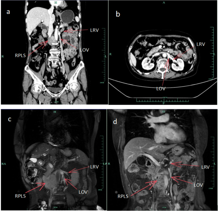
Case presentation: This case report describes a 75-year-old female patient who was admitted to the hospital with right lower abdominal pain and imaging studies suggesting a retroperitoneal mass and consideration of leiomyosarcoma. After the diagnosis was confirmed by imaging analysis combined with puncture biopsy, the patient received chemotherapy with adriamycin (75 mg/m²) and dacarbazine (200 mg/m²) to shrink the tumor once every 3 weeks for 4 cycles, which resulted in tumor shrinkage, and then underwent resection of the retroperitoneal leiomyosarcoma combined with resection of the inferior vena cava lesions. The retroperitoneal tumor involving the inferior vena cava (IVC) was completely resected without requiring IVC reconstruction during the surgical procedure. Final histopathological analysis confirmed the diagnosis of leiomyosarcoma. The patient demonstrated an uneventful postoperative recovery with no surgical complications, and subsequent follow-up evaluations revealed no evidence of disease recurrence, resulting in substantial improvement in the patient's quality of life on the basis of standardized oncology performance metrics.
Conclusions: This case emphasizes the critical role of imaging in diagnosis and surgical strategies. This study provides new ideas for the complete resection of retroperitoneal leiomyosarcoma. More studies are needed in the future to optimize the treatment strategy for this type of tumor.
Keywords: Inferior vena cava; Ovarian vein; Radiotherapy; Retroperitoneal leiomyosarcoma; Surgery.
© 2025. The Author(s).
Conflict of interest statement
Declarations. Ethics approval and consent to participate: This case report was approved by the Ethics Committee of the Affiliated People’s Hospital of Ningbo University and conducted according to the Helsinki and the IACUC guidelines. The above content can be reflected in the patient’s admission authorization letter without the need for an additional ethical code. Written informed consent was obtained from the patient for participation of this clinical case report. Consent for publication: Written informed consent was obtained from the patient for the publication of this case report and any accompanying images. Competing interests: The authors declare no competing interests.
Figures


References
-
- Ohmura Y, Takeda Y, Katsura Y, et al. [A case of retroperitoneal leiomyosarcoma with inferior Vena Cava invasion undergoing resection and reconstruction of the inferior Vena Cava and left renal vein]. Gan Kagaku Ryoho. 2024;51(13):1592–4. - PubMed
-
- Santoro R, Casciani E, Borrini F, Santoro E. Large retroperitoneal sarcoma invading the inferior Vena Cava successfully resected. Technical notes of two cases. Ann Ital Chir. 2023;94:404–10. - PubMed
LinkOut - more resources
Full Text Sources
Miscellaneous