. 2025 Sep 2;20(9):e0331303.
doi: 10.1371/journal.pone.0331303. eCollection 2025.

Mechanism of Huanglian Wendan Decoction in ameliorating non-alcoholic fatty liver disease via modulating gut microbiota-mediated metabolic reprogramming and activating the LKB1/AMPK pathway

Affiliations

Mechanism of Huanglian Wendan Decoction in ameliorating non-alcoholic fatty liver disease via modulating gut microbiota-mediated metabolic reprogramming and activating the LKB1/AMPK pathway

Jianping Zhu et al. PLoS One. .

Abstract

Background: Huanglian Wendan Decoction (HLWDD), a classical traditional Chinese medicine (TCM) formula, has shown therapeutic promise in treating metabolic disorders. However, its underlying mechanisms against non-alcoholic fatty liver disease (NAFLD) remain unclear.

Objective: This study aimed to elucidate the pharmacological mechanisms by which HLWDD ameliorates NAFLD, focusing on its impact on lipid metabolism, gut microbiota, and amino acid regulation.

Methods: A NAFLD rat model was established by administering a high-sugar, high-fat, high-salt diet for 20 weeks. The core components of HLWDD were identified and quantified using UPLC-Q-TOF-MS/MS and HPLC, and further validated via network pharmacology and molecular docking. Therapeutic efficacy was assessed through analysis of body weight, serum lipid profiles, inflammatory cytokines, hepatic histology, and protein expression. Gut microbiota composition and liver-intestine metabolite profiles were evaluated using metagenomic sequencing and LC-MS/MS.

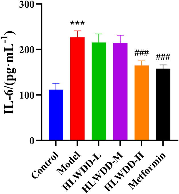
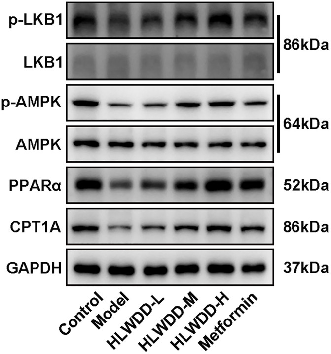
Results: Seven key constituents, including quercetin and berberine, were quantified (15.11-164.37 μg/mL) and shown to interact with lipid metabolism targets such as liver kinase B1 (LKB1), AMP-activated protein kinase (AMPK), peroxisome proliferator-activated receptor alpha (PPARα), and carnitine palmitoyltransferase 1A (CPT1A). HLWDD treatment significantly reduced body weight, hepatic lipid accumulation, and serum levels of triglycerides, total cholesterol, and low-density lipoprotein cholesterol, while increasing high-density lipoprotein cholesterol. Proinflammatory cytokines (IL-6, IL-1β, TNF-α) were notably suppressed. Mechanistically, HLWDD activated the LKB1/AMPK signaling pathway and modulated aspartic acid metabolism in association with increased abundance of Akkermansia in the gut. Metabolomic analysis identified 13 differential metabolites, with aspartic acid showing strong correlations with Akkermansia and LKB1/AMPK activity.

Conclusion: HLWDD exerts its anti-NAFLD effects by enhancing Akkermansia-mediated aspartate metabolism, thereby activating the LKB1/AMPK axis and promoting lipid oxidation via CPT1A and PPARα. This study provides new mechanistic insight into the gut-liver axis in NAFLD and highlights HLWDD as a multi-targeted therapeutic approach for restoring metabolic balance.


Conflict of interest statement

The authors declare no conflict of interest.

Figures

Fig 1
Fig 1. Analyses of chemical constituents of HLWDD using UPLC-Q-TOF-MS.
Base peak chromatogram of HLWDD in the negative-ion MS mode.
Fig 2
Fig 2. Analyses of chemical constituents of HLWDD using UPLC-Q-TOF-MS.
Base peak chromatogram of HLWDD in the positive-ion MS mode.
Fig 3
Fig 3. Network pharmacology analysis of HLWDD against NAFLD.
Diagram of the target intersection.
Fig 4
Fig 4. Network pharmacology analysis of HLWDD against NAFLD.
Interaction network diagram of core target proteins of HLWDD.
Fig 5
Fig 5. Network pharmacology analysis of HLWDD against NAFLD.
KEGG pathway enrichment results pathway diagram.
Fig 6
Fig 6. Network pharmacology analysis of HLWDD against NAFLD.
GO enrichment result bubble chart.
Fig 7
Fig 7. Network pharmacology analysis of HLWDD against NAFLD.
Drug–ingredients–genes–pathway interaction network diagram.
Fig 8
Fig 8. Molecular docking validation of HLWDD against NAFLD.
Representative docking results.
Fig 9
Fig 9. HPLC chromatograms of HLWDD.
Mixed reference substance. Peaks: 1. Quercetin, 2. Epiberberine, 3. Coptisine, 4. Palmatine, 5. Berberine, 6. Naringenin, 7. Houttuynia cordata.
Fig 10
Fig 10. HPLC chromatograms of HLWDD.
Sample extract. Peaks: 1. Quercetin, 2. Epiberberine, 3. Coptisine, 4. Palmatine, 5. Berberine, 6. Naringenin, 7. Houttuynia cordata.
Fig 11
Fig 11. HPLC chromatograms of HLWDD.
Blank control. Peaks: 1. Quercetin, 2. Epiberberine, 3. Coptisine, 4. Palmatine, 5. Berberine, 6. Naringenin, 7. Houttuynia cordata.
Fig 12
Fig 12. Therapeutic effects of HLWDD on NAFLD rats.
Body weight changes during intervention. Data expressed as mean ± SEM (n = 3). Statistical significance: **P < 0.01, ***P < 0.001 vs. Control; #P < 0.05, ##P < 0.01, ###P < 0.001 vs. Model.
Fig 13
Fig 13. Therapeutic effects of HLWDD on NAFLD rats.
Abdominal circumference changes during intervention. Data expressed as mean ± SEM (n = 3). Statistical significance: same as Fig 12.
Fig 14
Fig 14. Therapeutic effects of HLWDD on NAFLD rats.
Serum TC levels. Data expressed as mean ± SEM (n = 3). Statistical significance: same as Fig 12.
Fig 15
Fig 15. Therapeutic effects of HLWDD on NAFLD rats.
Serum TG levels. Data expressed as mean ± SEM (n = 3). Statistical significance: same as Fig 12.
Fig 16
Fig 16. Therapeutic effects of HLWDD on NAFLD rats.
Serum LDL-C levels. Data expressed as mean ± SEM (n = 3). Statistical significance: same as Fig 12.
Fig 17
Fig 17. Therapeutic effects of HLWDD on NAFLD rats.
Serum HDL-C levels. Data expressed as mean ± SEM (n = 3). Statistical significance: same as Fig 12.
Fig 18
Fig 18. Therapeutic effects of HLWDD on NAFLD rats.
Representative H&E staining of liver tissues (scale bar = 100 μm).
Fig 19
Fig 19. Therapeutic effects of HLWDD on NAFLD rats.
Serum IL-6 levels. Data expressed as mean ± SEM (n = 3). Statistical significance: same as Fig 12.
Fig 20
Fig 20. Therapeutic effects of HLWDD on NAFLD rats.
Serum IL-1β levels. Data expressed as mean ± SEM (n = 3). Statistical significance: same as Fig 12.
Fig 21
Fig 21. Therapeutic effects of HLWDD on NAFLD rats.
Serum TNF-α levels. Data expressed as mean ± SEM (n = 3). Statistical significance: same as Fig 12.
Fig 22
Fig 22. Effects of HLWDD on AMPK/PPARα signaling in rat liver.
Western blot bands of p-LKB1, LKB1, p-AMPK, AMPK, PPARα, CPT1A, and GAPDH.
Fig 23
Fig 23. Effects of HLWDD on AMPK/PPARα signaling in rat liver.
Relative protein expression of p-LKB1. Data are statistically presented as means ± SEM (n = 3). Compared with the Control, **P < 0.01, ***P < 0.001; compared with the Model, #P < 0.05, ##P < 0.01, ###P < 0.001.
Fig 24
Fig 24. Effects of HLWDD on AMPK/PPARα signaling in rat liver.
Relative protein expression of p-AMPK. Data expressed as mean ± SEM (n = 3). Statistical significance: same as Fig 23.
Fig 25
Fig 25. Effects of HLWDD on AMPK/PPARα signaling in rat liver.
Relative protein expression of PPARα. Data expressed as mean ± SEM (n = 3). Statistical significance: same as Fig 23.
Fig 26
Fig 26. Effects of HLWDD on AMPK/PPARα signaling in rat liver.
Relative protein expression of CPT1A. Data expressed as mean ± SEM (n = 3). Statistical significance: same as Fig 23.
Fig 27
Fig 27. Effects of HLWDD on AMPK/PPARα signaling in rat liver.
Relative mRNA expression of LKB1. Data expressed as mean ± SEM (n = 3). Statistical significance: same as Fig 23.
Fig 28
Fig 28. Effects of HLWDD on AMPK/PPARα signaling in rat liver.
Relative mRNA expression of AMPK. Data expressed as mean ± SEM (n = 3). Statistical significance: same as Fig 23.
Fig 29
Fig 29. Effects of HLWDD on AMPK/PPARα signaling in rat liver.
Relative mRNA expression of PPARα. Data expressed as mean ± SEM (n = 3). Statistical significance: same as Fig 23.
Fig 30
Fig 30. Effects of HLWDD on AMPK/PPARα signaling in rat liver.
Relative mRNA expression of CPT1A. Data expressed as mean ± SEM (n = 3). Statistical significance: same as Fig 23.
Fig 31
Fig 31. Multi-omics analysis of HLWDD-mediated gut–liver axis regulation in NAFLD.
Venn diagram of differential metabolites identified by gut metagenomics and liver metabolomics.
Fig 32
Fig 32. Multi-omics analysis of HLWDD-mediated gut–liver axis regulation in NAFLD.
Heatmap showing correlations between Akkermansia and key metabolites (aspartic acid, pantothenic acid).Color scales indicate Pearson correlation coefficients (r). Arrows highlight HLWDD-induced restorative trends. Data expressed as mean ± SEM (n = 3). Statistical significance: P < 0.05 (red), P < 0.01 (dark red).
Fig 33
Fig 33. Multi-omics analysis of HLWDD-mediated gut–liver axis regulation in NAFLD.
Gut microbiota–metabolite correlation network in model vs. HLWDD groups. Statistical significance: same as Fig 32.
Fig 34
Fig 34. Multi-omics analysis of HLWDD-mediated gut–liver axis regulation in NAFLD.
Hepatic metabolite correlation shifts (model vs. HLWDD-H). Statistical significance: same as Fig 32.
Fig 35
Fig 35. Multi-omics analysis of HLWDD-mediated gut–liver axis regulation in NAFLD.
Integrated heatmap of metabolite–protein interactions (LKB1/AMPK/CPT1A). Statistical significance: same as Fig 32.

Similar articles

References

    1. Harrison SA, Allen AM, Dubourg J, Noureddin M, Alkhouri N. Challenges and opportunities in NASH drug development. Nat Med. 2023;29(3):562–73. doi: 10.1038/s41591-023-02242-6 - DOI - PubMed
    1. Leow W-Q, Chan AW-H, Mendoza PGL, Lo R, Yap K, Kim H. Non-alcoholic fatty liver disease: the pathologist’s perspective. Clin Mol Hepatol. 2023;29(Suppl):S302–18. doi: 10.3350/cmh.2022.0329 - DOI - PMC - PubMed
    1. Grander C, Grabherr F, Tilg H. Non-alcoholic fatty liver disease: pathophysiological concepts and treatment options. Cardiovasc Res. 2023;119(9):1787–98. doi: 10.1093/cvr/cvad095 - DOI - PMC - PubMed
    1. Han SK, Baik SK, Kim MY. Non-alcoholic fatty liver disease: definition and subtypes. Clin Mol Hepatol. 2023;29(suppl):S5–16. doi: 10.3350/cmh.2022.0424 - DOI - PMC - PubMed
    1. Stern C, Castera L. Identification of high-risk subjects in nonalcoholic fatty liver disease. Clin Mol Hepatol. 2023;29(Suppl):S196–206. doi: 10.3350/cmh.2022.0431 - DOI - PMC - PubMed

MeSH terms

Substances