. 2025:5:10.3389/fddsv.2025.1545391.
doi: 10.3389/fddsv.2025.1545391. Epub 2025 Feb 20.

Transduction of hematopoietic stem and progenitor cells by an MECP2 lentiviral vector improves Rett syndrome phenotypes

Affiliations

Transduction of hematopoietic stem and progenitor cells by an MECP2 lentiviral vector improves Rett syndrome phenotypes

Joseph S Anderson et al. Front Drug Discov (Lausanne). 2025.

Abstract

Introduction: Rett Syndrome is a genetic neurodevelopmental disorder caused by decreased levels of MeCP2. Due to mutations in the MECP2 gene, insufficient MeCP2 protein levels lead to clinical phenotypes including the loss of normal movement, decreased communication, seizures, sleep disorders, and breathing problems. Currently there is no cure for Rett Syndrome and the only means to help patients is palliative care directed to their specific symptoms. Therefore, novel therapies need to be developed to alleviate disease phenotypes by restoring normal MECP2 expression. An autologous hematopoietic stem cell and gene therapy approach for Rett syndrome may offer a benefit to affected patients by systemic delivery of functional MeCP2, including to affected neurons in the central nervous system.

Methods: In our current experiments, we evaluated the therapeutic effect of MECP2 lentiviral vector transduced human CD34+ hematopoietic stem and progenitor cells after transplantation into an immunodeficient mouse model of Rett syndrome.

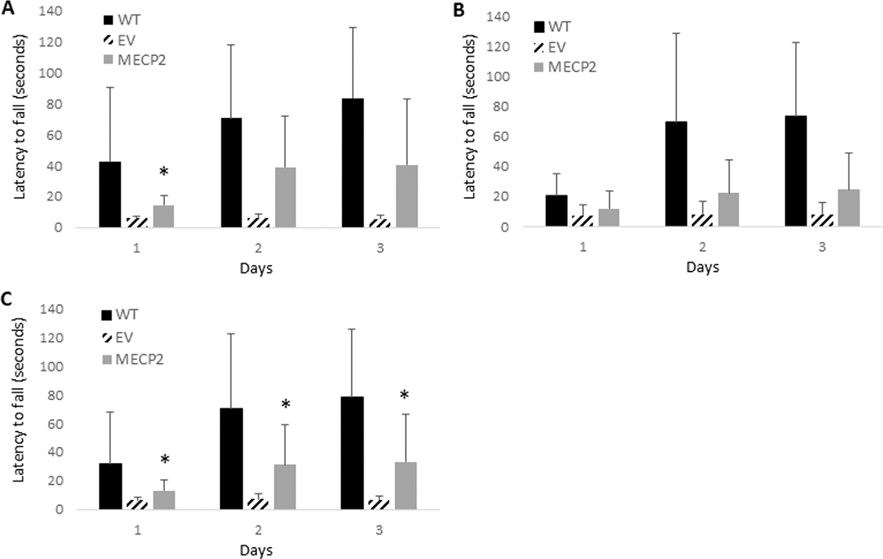
Results: We observed improvement of Rett syndrome-related phenotypes including the reversion toward normal motor abilities in an open field assay for total activity, horizontal activity, and vertical rearing activity, and an increased latency to fall in a rotarod assay. An increased level of MeCP2 protein was also observed in the brain tissue of transplanted mice.

Discussion: By providing functional MeCP2 to affected cells, our results highlight the ability of this strategy to improve Rett syndrome phenotypes. These proof-of-concept studies demonstrate the potential use of a stem cell gene therapy approach as a novel treatment for Rett syndrome patients.

Keywords: MECP2 lentiviral vector; Rett syndrome; gene therapy; hematopoietic stem and progenitor cells; neurodevelopmental disorders.


Conflict of interest statement

Conflict of interest The authors declare that the research was conducted in the absence of any commercial or financial relationships that could be construed as a potential conflict of interest. The author(s) declared that they were an editorial board member of Frontiers, at the time of submission. This had no impact on the peer review process and the final decision.

Figures

FIGURE 1
FIGURE 1
Functionality of an MECP2 expressing lentiviral vector: (A) The modified MECP2 gene was cloned into a third-generation self-inactivating lentiviral vector. The control empty vector (EV) consists of the same vector backbone without the MECP2 transgene. (B) Rett syndrome-affected B lymphocytes were left nontransduced (NT) or transduced with the EV or MECP2 vector. Total RNA was extracted and MECP2 transcripts were quantified by QPCR (N = 3). Data is presented as a fold-increase in expression normalized to 1.0 from the NT data. Mean and standard error of the mean are shown. (C) Total protein was extracted from the cells and evaluated for expression of MeCP2 (~75 kDa) by Western blot. GAPDH (~25 kDa) was used as an internal loading control. *p < 0.001.
FIGURE 2
FIGURE 2
CFU assay, cell expansion, and expression of MECP2 in human CD34+ derived macrophages: Human CD34+ HSPC were left nontransduced (NT) or transduced with the empty vector (EV) or the MECP2 vector and cultured in a CFU assay with methylcellulose media. (A) Total GM, GEMM, and BFU-E colonies were counted. (B) Total cell expansion was evaluated by comparing the number of input cells to the number of cells after the CFU assay. (C) Total RNA was extracted from the CD34+ derived macrophages post-CFU assay and MECP2 transcripts were quantified by QPCR. Data is presented as a fold-increase in expression normalized to 1.0 from the NT data. Mean and standard error of the mean are shown. *p < 0.01.
FIGURE 3
FIGURE 3
Rotarod analysis of BRM mice transplanted with MECP2 vector transduced human CD34+ HSPC: BRM mice were transplanted with human CD34+ HSPC transduced with either the empty vector (EV) or the MECP2 vector MECP2). Nontransplanted MECP2 wild type (WT) (B6-Rag2−/− Mecp2+/+) mice were used as controls. Sixteen weeks post-transplant, the mice were subjected to a rotarod assay for three consecutive days. Total latency time to fall was measured with a maximum time of 5 min. Data displayed from transplanted (A) newborn mice (N = 4), (B) adult mice (N = 4), or (C) a combination of all mice (N = 8). Mean and standard error of the mean are shown. *p < 0.05.
FIGURE 4
FIGURE 4
Open field analysis of BRM mice transplanted with MECP2 vector transduced human CD34+ HSPC: BRM mice were transplanted with human CD34+ HSPC transduced with either the empty vector (EV) or the MECP2 vector (MECP2). Nontransplanted MECP2 wild type (WT) (B6-Rag2−/− Mecp2+/+) mice were used as controls. Sixteen weeks post-transplant, the mice were subjected to a 15-minute open field assay to evaluate (A) total distance traveled, (B) total horizontal activity, and (C) vertical rearing activity. Data is displayed from transplanted newborn mice (N = 4), adult mice (N = 4), or a combination of all mice (N = 8). Mean and standard error of the mean are shown. *p < 0.05 and **p < 0.01.
FIGURE 5
FIGURE 5
Immunohistochemical detection of MECP2 in brain tissue obtained from BRM mice: Sagittal sections of brain tissue obtained from the nontransplanted wild type (WT), empty vector (EV) transduced, or MECP2 expressing (MECP2) vector transduced cell transplanted mice were labeled with a monoclonal antibody for mouse MECP2 (N = 7). Enumeration of positive cells was performed and compared between the groups. Data displayed from transplanted (A) newborn mice, (B) adult mice, or (C) a combination of all mice. (D) Representative stained images from the brain tissues are displayed for the WT, EV, and MECP2 groups with arrows indicating positive cells. The scale bar in the upper left represents 50 μm. Mean and standard error of the mean are shown. *p = 0.05 **p = 0.03.

References

    1. Adhikari A, Copping NA, Beegle J, Cameron DJ, Deng P, O’Geen H, et al. (2021). Functional rescue in an Angelman syndrome model following treatment with lentivector transduced hematopoietic stem cells. Hum. Mol. Genet. 30 (12), 1067–1083. doi: 10.1093/hmg/ddab104 - DOI - PMC - PubMed
    1. Alvarez-Saavedra M, Saez MA, Kang D, Zoghbi HY, and Young JI (2007). Cell-specific expression of wild-type MeCP2 in mouse models of Rett syndrome yields insight about pathogenesis. Hum. Mol. Genet. 16 (19), 2315–2325. doi: 10.1093/hmg/ddm185 - DOI - PubMed
    1. Anderson JS, Lodigiani AL, Barbaduomo CM, and Beegle JR (2024). Hematopoietic stem cell gene therapy for the treatment of SYNGAP1-related non-specific intellectual disability. J. Gene Med 26 (7), e3717. doi: 10.1002/jgm.3717 - DOI - PubMed
    1. Beegle J, Hendrix K, Maciel H, Nolta JA, and Anderson JS (2020). Improvement of motor and behavioral activity in Sandhoff mice transplanted with human CD34+ cells transduced with a HexA/HexB expressing lentiviral vector. J. Gene Med. 22 (9), e3205. doi: 10.1002/jgm.3205 - DOI - PMC - PubMed
    1. Ben-Shachar S, Chahrour M, Thaller C, Shaw CA, and Zoghbi HY (2009). Mouse models of MeCP2 disorders share gene expression changes in the cerebellum and hypothalamus. Hum. Mol. Genet. 18 (13), 2431–2442. doi: 10.1093/hmg/ddp181 - DOI - PMC - PubMed

LinkOut - more resources