. 2025 Aug 16:34:102210.
doi: 10.1016/j.mtbio.2025.102210. eCollection 2025 Oct.

Phosphorus-32 microspheres: A dual-modality transarterial radioembolization approach for hepatocellular carcinoma therapy and Anti-PD1 immunotherapy potentiation

Affiliations

Phosphorus-32 microspheres: A dual-modality transarterial radioembolization approach for hepatocellular carcinoma therapy and Anti-PD1 immunotherapy potentiation

Shipeng Dai et al. Mater Today Bio. .

Abstract

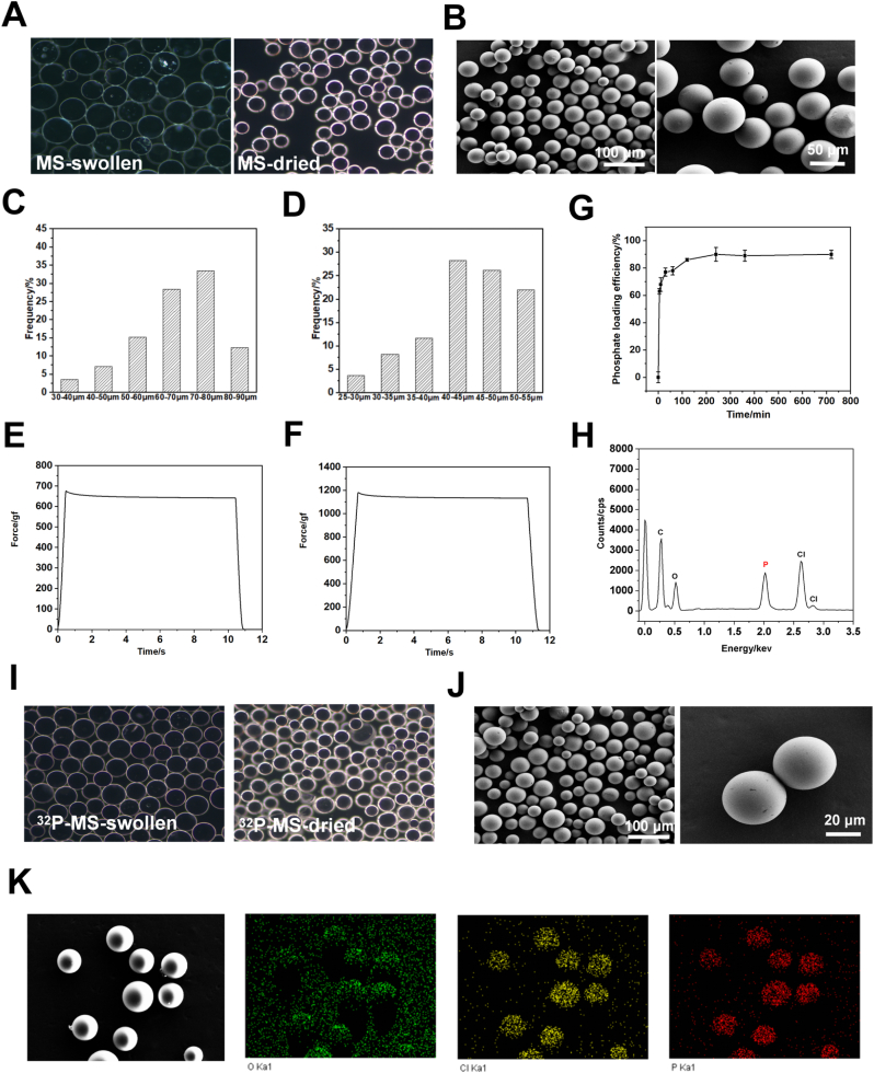
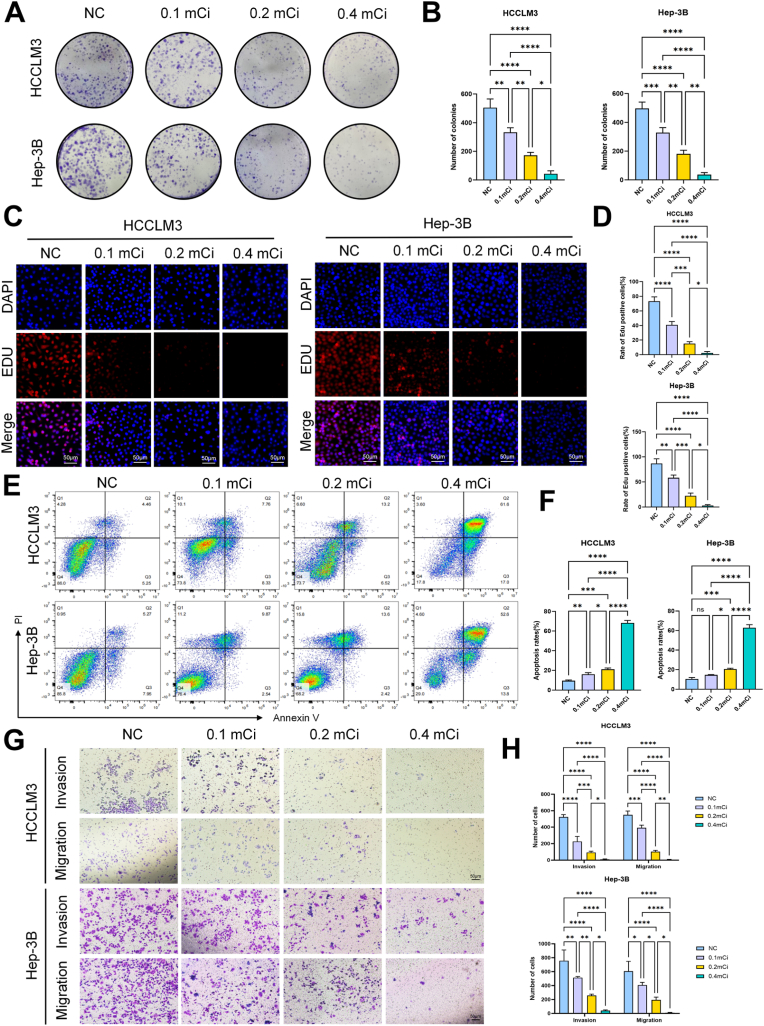
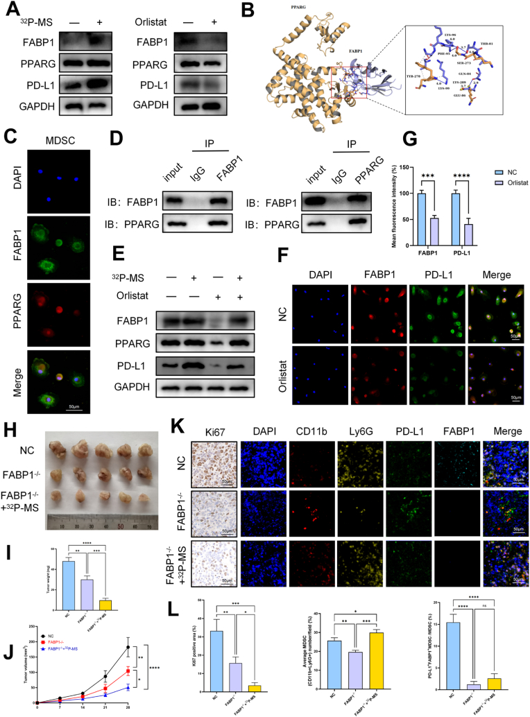
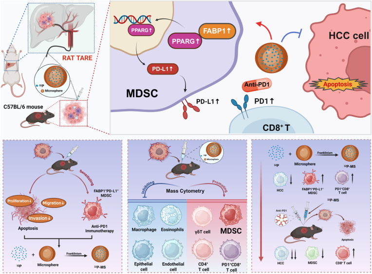
Transarterial radioembolization (TARE) is a key therapy for hepatocellular carcinoma (HCC) management and downstaging. While 90Y microspheres (glass/resin) are widely used, their clinical application is limited by complexity, short half-life, and high costs. Thus, novel radionuclide microspheres are crucial. This study developed phosphorus-32-loaded microspheres (32P-MS). In vitro, 32P-MS dose-dependently suppressed HCC cell proliferation, migration, and invasion while inducing apoptosis. In vivo, 32P-MS TARE achieved tumor vascular embolization, reducing tumor vol/wt (confirmed by Positron Emission Tomography-Computed Tomography (PET-CT), Hematoxylin and Eosin (HE) staining, TUNEL/Ki67 assays without systemic toxicity. RNA sequencing and mass cytometry analyses revealed 32P-MS upregulated FABP1+PD-L1+ myeloid-derived suppressor cell (MDSC), linked to immunosuppression. Mechanistic investigations, including molecular docking, co-localization, and co-immunoprecipitation (Co-IP) assays, demonstrated that 32P-MS activated the FABP1/PPARG/PD-L1 axis in MDSC. Genetic ablation of FABP1 or pharmacological inhibition with Orlistat reversed PD-L1 expression and augmented anti-tumor efficacy. Combining 32P-MS with anti-PD1 therapy synergistically suppressed tumor growth, reduced MDSC infiltration, and reinvigorated CD8+ T cell activity, significantly improving treatment sensitivity. 32P-MS is a promising HCC therapeutic with dual anti-tumor and immune-modulatory functions, providing a compelling rationale for integrating radioembolization with immune checkpoint blockade to counteract immunosuppressive resistance in HCC.

Keywords: Hepatocellular carcinoma; Myeloid-derived suppressor cell; PD1; Phosphorus-32-loaded microspheres; Transarterial radioembolization.


Conflict of interest statement

The authors declare that they have no competing interests.

Figures

Image 1
Graphical abstract
Fig. 1
Fig. 1
Characterization and drug delivery capacity of microspheres (A) Optical micrographs of microspheres-swollen, microspheres-dried. (B) SEM of microspheres. (C- D)Particle size distribution of microspheres-swollen (C), microspheres-dried (D). (E-F)Time-force curves of 50 % strain (E) and 80 % strain (F) compressed microspheres under texture analysis. (G) Loading curve of phosphate ion. (H) The element analysis of 32P-MS. (I) Optical micrographs 32P-MS-swollen, 32P-MS-dried. (J) SEM of 32P-MS. (K) chlorine, oxygen and phosphorus element mapping images of 32P-MS.
Fig. 2
Fig. 2
32P-MS inhibits the proliferation, invasion and migration of HCC in vitro (A-B) Colony formation assay was carried out to evaluate the proliferation and colony formation ability of HCC cells treated with different doses of 32P-MS radiation. (C–D) EdU assay was carried out to evaluate the proliferation of HCC cells treated with different doses of 32P-MS radiation; the scale bar represents 50 μm. (E–F) Apoptosis in HCC cells across various treatments was gauged with flow cytometry. (G–H) Transwell assay was used to assess the invasion and migration ability of HCC cells treated with different doses of 32P-MS radiation; the scale bar represents 50 μm. (ns = not significant, ∗P < 0.05, ∗∗P < 0.01, ∗∗∗P < 0.001, ∗∗∗∗P < 0.0001).
Fig. 3
Fig. 3
TARE with 32P-MS shows good embolization performance and significantly inhibits tumor growth in SD rat HCC model (A) Real-time DSA of pre- and post-embolizing in 32P-MS radiation group. (B) ECT showed that the nuclides were mainly concentrated in the liver. (C) Transverse section, coronal section and median sagittal section of rats' PET-CT images before TACE and 7 days after TACE in 32P-MS radiation group. (D) Overview of rat tumors and HE staining picture of respective cancer tissues treated with 32P-MS radiation or not (scale bars, 50 μm). (E–F) IF staining to detect the expression of TUNEL in rat HCC tissues treated with 32P-MS radiation or not (scale bars, 50 μm). (G–H) IF staining to detect the expression of Ki67 in rat HCC tissues treated with 32P-MS radiation or not (scale bars, 50 μm). (I) HE staining picture of respective rat organs or tissues treated with 32P-MS radiation or not (scale bars, 50 μm). (∗P < 0.05, ∗∗P < 0.01, ∗∗∗P < 0.001, ∗∗∗∗P < 0.0001).
Fig. 4
Fig. 4
32P-MS inhibits tumor growth compared with C57BL/6 mouse HCC models and shows a good biosafety (A) Overview of mice subcutaneous tumors and HE staining picture of respective cancer tissues treated with 32P-MS radiation or not (scale bars, 50 μm). (B–C) Tumor growth curves and tumor weights of each group. (D–E) IF staining to detect the expression of TUNEL in mice HCC tissues treated with 32P-MS radiation or not (scale bars, 50 μm). (F–G) IF staining to detect the expression of Ki67 in mice HCC tissues treated with 32P-MS radiation or not (scale bars, 50 μm). (H) HE staining picture of respective mice organs or tissues treated with 32P-MS radiation or not (scale bars, 50 μm). (I) Representative a-SMA Immunohistochemistry of livers (scale bars, 50 μm). (ns = not significant, ∗p < 0.05, ∗∗p < 0.01, ∗∗∗p < 0.001).
Fig. 5
Fig. 5
RNA sequencing and mass cytometry results after 32P-MS treatment (A- B) The volcano map and radar map showed differentially expressed genes between cancer tissues from control group or 32P-MS radiation group. (C–D) Pathway enrichment of KEGG among the diverse genes expressed. (E) Tumor samples from control group or 32P-MS radiation group were detected subcutaneous by mass cytometry. Single, viable, and intact CD45+ immune cells were recycled from selected cells in the respective tissues. CD45+ immune cell aggregation and subgroup annotation were observed in all samples. In total, there were 41 cell clusters. (F–H) The TSNE scatter plot showed the distribution of each cell cluster, and the proportion of each cell cluster was statistically analyzed. (I) The histogram showing the number of the respective cell cluster in different groups by mass cytometry.
Fig. 6
Fig. 6
32P-MS therapy results in an increased number of FABP1+PD-L1+ MDSC in HCC (A- B) The UMAP plot demonstrated that 10 common immune cell clusters were named according to marker identification and merger. (C) The bar chart illustrated the marking identification and merging process that considered in the naming of 10 samples. (D) The bar chart illustrated the proportion of different immune cell clusters in HCC tissues. (E–F) Myeloid cells were mainly divided into 6 cell populations according to marker gene expression. (G) The enrichment of marker genes in myeloid cells. (H–I) The expression of MDSC surface markers CD11b, Ly6G increased in the 32P-MS radiation group relative to the control group, the expression of FABP1, and CD274 (PD-L1) increased in MDSC in the 32P-MS radiation group relative to the control group. (J) Representative CD11b, Ly6G, FABP1, and CD274 fluorescence immunohistochemical of tumors (scale bars, 50 μm). n = 3 mice per group. (∗∗ p < 0.01, ∗∗∗∗ p < 0.0001).
Fig. 7
Fig. 7
FABP1 promotes the expression of PD-L1 by regulating PPARG, thus maintaining the function of MDSC in HCC (A) Protein expression levels of FABP1, PPARG and PD-L1 in human MDSC after treated with 32P-MS radiation, Orlistat or not. GAPDH is used for loading control. (B) Artificial intelligence (AI) technology was used to docking and found that FABP1 and PPARG proteins may also have action sites. (C) Representative FABP1, PPARG Immunohistochemistry of human MDSC. (D) Co‐IP and western blot analysis showing the interaction of FABP1 and PPARG in human MDSC. (E) Western blot analysis was used to verify that Orlistat intervention could partially reverse the upregulation of FABP1 and PPARG proteins in human MDSC induced by 32P-MS radiation. (F–G) Representative FABP1, PD-L Immunohistochemistry of human MDSC treated with Orlistat or not. (E) Photographs of tumors induced by the subcutaneous inoculation of mice (n = 5 mice per group) with transfected hepa1-6 cells. (F) Graphs of tumor weights. n = 5 mice per group. (G) Growth curves of tumor volumes. n = 5 mice per group. (K–L) Representative Ki67 Immunohistochemistry of tumors (scale bars, 50 μm). Representative CD11b, Ly6G, FABP1, and CD274 fluorescence immunohistochemical of tumors (scale bars, 50 μm). (ns = not significant, ∗ p < 0.05, ∗∗ p < 0.01, ∗∗∗ p < 0.001, ∗∗∗∗ p < 0.0001).
Fig. 8
Fig. 8
32P-MS significantly enhances the sensitivity of anti-PD1 in HCC treatment (A) Graphs of tumor weights. n = 5 mice per group. (B) Growth curves of tumor volumes. n = 5 mice per group. (C) Representative Ki67 fluorescence immunohistochemical of tumors (scale bars, 50 μm). (D) Representative CD8, PD1 fluorescence immunohistochemical of tumors (scale bars, 50 μm). (E) Representative CD11b, Ly6G fluorescence immunohistochemical of tumors (scale bars, 50 μm). (F) Statistical analysis of (C–E). (ns = not significant, ∗ p < 0.05, ∗∗ p < 0.01, ∗∗∗ p < 0.001, ∗∗∗∗ p < 0.0001).
Fig. 9
Fig. 9
Pattern diagram. We developed 32P-MS for TARE in HCC. 32P-MS demonstrated robust anti-tumor efficacy, including dose-dependent suppression of HCC proliferation, migration, and invasion in vitro, alongside significant tumor vascular embolization and growth inhibition in vivo across rodent models. Mechanistically, 32P-MS uniquely modulated the immunosuppressive tumor microenvironment by upregulating FABP1+PD-L1+MDSC through activation of the FABP1/PPARG/PD-L1 axis. Importantly, combining 32P-MS with anti-PD1 therapy amplified anti-tumor responses by reducing MDSC infiltration, revitalizing CD8+ T cell activity, and overcoming immunosuppressive resistance.

Similar articles

References

    1. Siegel R.L., Miller K.D., Jemal A. Cancer statistics. CA Cancer J. Clin. 2019;69(1):7–34. 2019. - PubMed
    1. Bruix J., Sherman M., D. American Association for the Study of Liver Management of hepatocellular carcinoma: an update. Hepatology. 2011;53(3):1020–1022. - PMC - PubMed
    1. Riaz A., et al. Imaging response in the primary index lesion and clinical outcomes following transarterial locoregional therapy for hepatocellular carcinoma. JAMA. 2010;303(11):1062–1069. - PMC - PubMed
    1. Saini A., et al. History and evolution of Yttrium-90 radioembolization for hepatocellular carcinoma. J. Clin. Med. 2019;8(1) - PMC - PubMed
    1. Salem R., et al. Y90 radioembolization significantly prolongs time to progression compared with chemoembolization in patients with hepatocellular carcinoma. Gastroenterology. 2016;151(6):1155–1163 e2. - PMC - PubMed

LinkOut - more resources