

doi: 10.21037/qims-2025-176.
Epub 2025 Aug 11.
Endoscopic endonasal transsphenoidal approach of pituitary macroadenoma and optic canal stenosis in a patient with McCune-Albright syndrome
Affiliations
- PMID: 40893547
- PMCID: PMC12397750
- DOI: 10.21037/qims-2025-176
Item in Clipboard
Endoscopic endonasal transsphenoidal approach of pituitary macroadenoma and optic canal stenosis in a patient with McCune-Albright syndrome
Quant Imaging Med Surg.
.
No abstract available
Keywords: McCune-Albright; bone dysplasia; optic canal; pituitary macroadenoma.
Conflict of interest statement
Conflicts of Interest: All authors have completed the ICMJE uniform disclosure form (available at https://qims.amegroups.com/article/view/10.21037/qims-2025-176/coif). The authors have no conflicts of interest to declare.
Figures
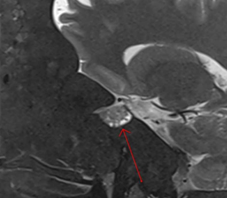
Sagittal T2-weighted MRI illustrating a pituitary macroadenoma in the patient (red arrow). MRI, magnetic resonance imaging.

Axial tomography imaging of facial bones. There is considerable osseous enlargement with regions of varied bone density ranging from sclerotic to lucent. The majority of affected bones exhibit a ground-glass appearance.

Axial T2-weighted MRI. Notice the involvement of the optic canal (red arrows). MRI, magnetic resonance imaging.

Coronal T1-weighted Gd MRI illustrating a pituitary macroadenoma touching the optic chiasm, with left parasellar extension (left side). Gd, gadolinium; MRI, magnetic resonance imaging.

Intraoperative endoscopic view demonstrating a narrowed left nasal fossa. *, indicates inferior turbinate; #, indicates nasal septum.
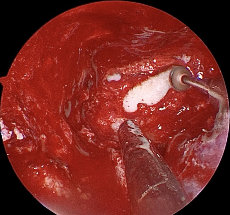
Endoscopic intraoperative image depicting the surgical drilling of the extensively ossified sphenoid sinus.

Endoscopic intraoperative images illustrating the exposure of the dura mater overlying the sellar region and the optic canal, following drilling of the sphenoid sinus (A); the initiation of pituitary adenoma resection (B) and the final aspect of the sellar region after gross total resection of the pituitary adenoma (C). *, indicates optic canal; #, indicates sellar region.

Endoscopic intraoperative image illustrating the exposure of the dura mater overlying the sellar region and the optic canal, following drilling of the sphenoid sinus. *, indicates optic canal; #, indicates sellar region.

Endoscopic intraoperative image showing the initiation of pituitary adenoma resection.

Endoscopic intraoperative image showing the final aspect of the sellar region after gross total resection of the pituitary adenoma.
References
-
- Albright F, Butler AM, Hampton AO, Smith PH. Syndrome characterized by osteitis fibrosa disseminata, areas of pigmentation and endocrine dysfunction, with precocious puberty in females: report of five cases. N Engl J Med 1937;216:727-46.
-
- Sammut SJ, Kandasamy J, Newman W, Sinha A, Ross J, Blair JC, May P. Relief of severe retro-orbital pain and vision improvement after optic-nerve decompression in polyostotic fibrous dysplasia: case report and review of the literature. Childs Nerv Syst 2008;24:515-20. 10.1007/s00381-007-0543-y - DOI - PubMed
Publication types
LinkOut - more resources
Full Text Sources