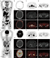
Comparison of [68Ga]Ga-FAPI-04 and [18F]FDG PET/CT for detection of bone metastases of lung cancer
- PMID: 40893562
- PMCID: PMC12397704
- DOI: 10.21037/qims-2025-234
Comparison of [68Ga]Ga-FAPI-04 and [18F]FDG PET/CT for detection of bone metastases of lung cancer
Abstract
Background: Bone metastases of lung cancer typically indicate disease progression and poor prognosis. Early and accurate detection is crucial for staging, treatment planning, and prognostic evaluation. This study aimed to compare the diagnostic value of gallium 68-labeled fibroblast-activation protein inhibitor-04 ([68Ga]Ga-FAPI-04) and fluorine 18-labeled fluorodeoxyglucose ([18F]FDG) positron-emission tomography/computed tomography (PET/CT) imaging in detecting bone metastases in lung cancer.
Methods: A retrospective analysis was conducted on patients with pathologically confirmed lung cancer and clinically suspected bone metastases. These patients underwent both [68Ga]Ga-FAPI-04 and [18F]FDG PET/CT imaging. Initially, all patient images were visually evaluated, and the diagnostic efficacy of the two imaging methods was compared at both the patient and lesion levels for detecting bone metastases from lung cancer. Additionally, a semi-quantitative analysis was performed to compare the optimal maximum standardized uptake value (SUVmax) threshold and diagnostic efficacy of the two examinations for diagnosing benign and malignant bone lesions.
Results: A total of 25 lung cancer patients were included in the study, with nine confirmed cases and 133 lesions of bone metastases. At the patient level, there were no statistically significant differences in the detection rate, sensitivity, specificity, positive predictive value, negative predictive value, or accuracy between [68Ga]Ga-FAPI-04 and [18F]FDG PET/CT for identifying patients with bone metastases (P>0.05). At the lesion level, the detection rate, sensitivity, negative predictive value, and accuracy of [68Ga]Ga-FAPI-04 PET/CT for detecting bone metastases were higher than those of [18F]FDG PET/CT (81.37% vs. 57.14%, 98.50% vs. 69.17%, 88.24% vs. 34.92%, 90.68% vs. 70.81%), with statistically significant differences (P<0.01). The SUVmax of malignant bone lesions on both [68Ga]Ga-FAPI-04 and [18F]FDG PET/CT was significantly higher than those of benign bone lesions, with statistically significant differences (P<0.05). Moreover, the SUVmax of benign and malignant bone lesions on [68Ga]Ga-FAPI-04 PET/CT was significantly higher than those on [18F]FDG PET/CT, with statistically significant differences (P<0.01). In [68Ga]Ga-FAPI-04 and [18F]FDG PET/CT imaging, the area under the curves (AUCs) of SUVmax for diagnosing bone metastases were 0.856 and 0.724, respectively, with statistically significant differences (P<0.05); the optimal diagnostic thresholds were 5.38 and 3.77, respectively. The sensitivity, negative predictive value, and accuracy of SUVmax based on [68Ga]Ga-FAPI-04 PET/CT for diagnosing lung cancer bone metastases were higher than those based on [18F]FDG PET/CT (80.45% vs. 65.26%, 46.49% vs. 23.26%, 81.25% vs. 67.29%), with statistically significant differences (P<0.05).
Conclusions: Compared to [18F]FDG PET/CT, [68Ga]Ga-FAPI-04 PET/CT significantly improves the detection rate of lung cancer bone metastases at the lesion level. Additionally, [68Ga]Ga-FAPI-04 PET/CT offers superior image contrast and higher SUVmax, which also contribute to improving the accuracy of lung cancer bone metastasis diagnosis. This allows for more accurate staging of patients, enabling precise individualized treatment and improving patient prognosis.
Keywords: Bone metastasis; computed tomography (CT); fibroblast-activation protein inhibitor (FAPI); fluorodeoxyglucose (FDG); positron-emission tomography (PET).
Copyright © 2025 AME Publishing Company. All rights reserved.
Conflict of interest statement
Conflicts of Interest: All authors have completed the ICMJE uniform disclosure form (available at https://qims.amegroups.com/article/view/10.21037/qims-2025-234/coif). The authors have no conflicts of interest to declare.
Figures


Similar articles
-
Diagnostic Value of [18F]-FDG and [68 Ga]-FAPI-04 PET/MRI for Lymph Node Metastasis in Papillary Thyroid Cancer.Mol Imaging Biol. 2025 Aug;27(4):540-549. doi: 10.1007/s11307-025-02028-x. Epub 2025 Jun 24. Mol Imaging Biol. 2025. PMID: 40555936
-
123I-MIBG scintigraphy and 18F-FDG-PET imaging for diagnosing neuroblastoma.Cochrane Database Syst Rev. 2015 Sep 29;2015(9):CD009263. doi: 10.1002/14651858.CD009263.pub2. Cochrane Database Syst Rev. 2015. PMID: 26417712 Free PMC article.
-
68Ga-FAPI and 18F-FAPI PET/CT for detection of nodal metastases prior radical cystectomy in high-risk urothelial carcinoma patients.Eur J Nucl Med Mol Imaging. 2025 Sep;52(11):3963-3974. doi: 10.1007/s00259-025-07239-6. Epub 2025 Apr 24. Eur J Nucl Med Mol Imaging. 2025. PMID: 40272498 Free PMC article.
-
Head-To-Head Comparison of 68Ga-FAPI PET/CT and FDG PET/CT for the Detection of Peritoneal Metastases: Systematic Review and Meta-Analysis.AJR Am J Roentgenol. 2023 Apr;220(4):490-498. doi: 10.2214/AJR.22.28402. Epub 2022 Nov 2. AJR Am J Roentgenol. 2023. PMID: 36321984
-
Fluorine-18-fluorodeoxyglucose (FDG) positron emission tomography (PET) computed tomography (CT) for the detection of bone, lung, and lymph node metastases in rhabdomyosarcoma.Cochrane Database Syst Rev. 2021 Nov 9;11(11):CD012325. doi: 10.1002/14651858.CD012325.pub2. Cochrane Database Syst Rev. 2021. PMID: 34753195 Free PMC article.
References
-
- Forrai G, Kovács E, Ambrózay É, Barta M, Borbély K, Lengyel Z, Ormándi K, Péntek Z, Tünde T, Sebő É. Use of Diagnostic Imaging Modalities in Modern Screening, Diagnostics and Management of Breast Tumours 1st Central-Eastern European Professional Consensus Statement on Breast Cancer. Pathol Oncol Res 2022;28:1610382. 10.3389/pore.2022.1610382 - DOI - PMC - PubMed
LinkOut - more resources
Full Text Sources
