. 2025 Jul 22;4(4):363-377.
doi: 10.1002/mlf2.70018. eCollection 2025 Aug.

Mechanistic insights into the T6SS of multi-drug-resistant Aeromonas hydrophila and its role in competition and pathogenesis

Affiliations

Mechanistic insights into the T6SS of multi-drug-resistant Aeromonas hydrophila and its role in competition and pathogenesis

Hao Wang et al. mLife. .

Abstract

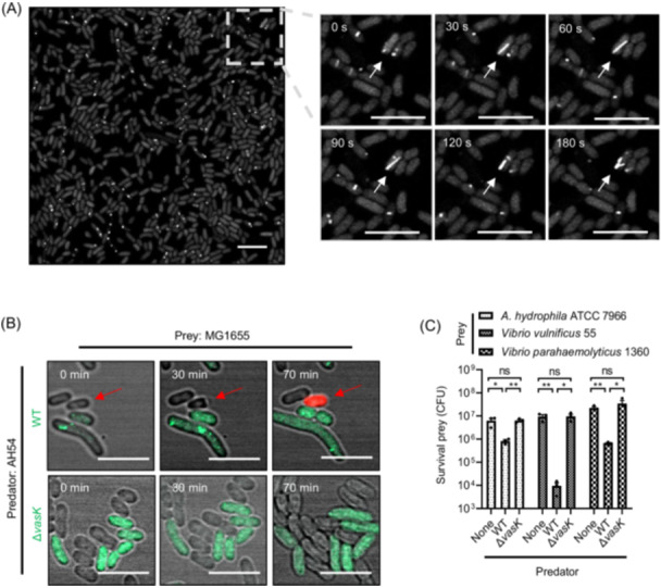
Aeromonas hydrophila, an opportunistic pathogen, often encodes Type VI Secretion System (T6SS) genes. However, the specific functions of T6SS, particularly in the context of clinical strains, remain poorly understood. In this study, we characterize a multi-drug-resistant strain, AH54, which possesses a complete and functional T6SS, composed of a structural cluster and two homologous auxiliary clusters (Aux1 and Aux2). Each auxiliary cluster encodes two distinct effector proteins: a rearrangement hotspot (Rhs) protein and a proline-alanine-arginine repeat (PAAR) protein-Rhs1/PAAR1 in Aux1 and Rhs2/PAAR2 in Aux2. Our findings reveal that AH54 assembles a fully operational T6SS capable of delivering these effectors, driving inter-bacterial antagonism. Interestingly, the T6SS activity in AH54 is temperature-regulated, with enhanced secretion and antibacterial activity at lower temperatures. To protect itself from self-intoxication, AH54 produces immunity proteins (Tsi1-Tsi4) that neutralize the toxic effectors. While PAAR1 and PAAR2 are critical for Hcp secretion, immunity proteins Tsi3 and Tsi4 do not cross-protect against PAAR effectors, suggesting distinct roles for each PAAR protein in optimizing AH54's competitive fitness. In addition, using a Dictyostelium discoideum phagocytosis model, we demonstrate that Rhs2, a metal ion-dependent DNase effector, plays a crucial role in protecting AH54 from eukaryotic predation via T6SS. These findings highlight the pivotal role of T6SS in bacterial competition and pathogenesis, offering new insights into the virulence mechanisms of A. hydrophila.

Keywords: Aeromonas species; T6SS; anti‐eukaryotic virulence; microbial interaction.


Conflict of interest statement

The authors declare no conflict of interests.

Figures

Figure 1
Figure 1
Genetic characterizations of T6SS gene clusters in Aeromonas hydrophila strains. (A–C) Comparative genomics analysis of the T6SS structural cluster (A), auxiliary cluster I (B) and cluster II (C) in A. hydrophila AH54, ATCC 7966, and NJ‐35. Four hypothetical effector–immunity (E–I) protein pairs from the A. hydrophila AH54 were identified and displayed. These E–I pairs are highlighted in red (effector) and blue (immunity). Sequence alignment was performed using BLASTn and the conserved sequence was visualized using EasyFig. (D, E) Schematic domain organization of T6SS effector proteins Rhs1/Rhs2 (D) and PAAR1/PAAR2 (E). Conserved domains were predicted using Batch CD‐Search.
Figure 2
Figure 2
Functional characterizations of T6SS in A. hydrophila clinical strain AH54. (A) T6SS dynamics in AH54. VipA_sfGFP localization (sheath assembly) was monitored in at least 200 AH54 vipA_sfGFP cells by time lapse microscopy. Representative images of the GFP fluorescence channel are shown. Additional images with the time series of view can be found in Movie S1. The white arrow indicates the assembly and depolymerization of the T6SS during 180 s. Scale bar, 5 μm. (B) Competitive killing assay of predator AH54 and prey MG1655. AH54 vipA_sfGFP wild‐type (WT, upper) or its T6SS‐deficient mutant ΔvasK by knockout of an allelic T6SS gene (lower) was mixed with MG1655 at a 2 :1 ratio and blotted on a LB agar plate with propidium iodide (PI) dye at 28°C. Green cells indicate predator, cells without fluorescence represent prey, and red cells indicate dead prey cell during T6SS‐meidated competition (red arrow). A representative view from three similar results is displayed. The data of successive photos were related to Movies S2 and S3. Scale bar, 5 μm. (C) Bacterial competition assays between A. hydrophila and closely related species. Quantification of survival prey A. hydrophila ATCC 7966, V. vulnificus 55, and V. parahaemolyticus 1360 after a T6SS attack by predator AH54 WT and ΔvasK. The values represent mean ± SEM of three independent experiments. Statistical significance was determined using an unpaired Student's t‐test (ns, nonsignificant; *p < 0.05; **p < 0.01).
Figure 3
Figure 3
Temperature affects T6SS dynamics in AH54. (A) T6SS sheaths of AH54 vipA_sfGFP overnight cultures grown at 22°C, 28°C, and 37°C and monitored by confocal microscopy. Representative fluorescence micrographs from three independent replicates are shown. Scale bar, 5 μm. (B) Quantification of the sheath numbers per AH54 cell grown at 22°C, 28°C, and 37°C. The bold horizontal bar represents the median value; the bottom and top dash line of the internal boxplot correspond to the 25th and 75th percentiles, respectively. p‐values from all data were determined using an unpaired Student's t‐test, and statistical significance is indicated above the plots (ns, nonsignificant; ****p < 0.0001). (C) Western blot analysis of Hcp secretion in AH54 at different temperatures. AH54 was grown in LB medium to an OD600 of 1.5 at 22°C, 28°C, and 37°C. Cell pellets and 4 ml supernatants (Sup) were analyzed by SDS‐PAGE and immunoblot assays using anti‐Hcp and anti‐EF‐Tu primary antibodies. (D) Effect of temperature on AH54 T6SS‐mediated competition. A mixture of predator (AH54 WT or ΔvasK) and prey (MG1655) was grown in LB medium at indicated temperatures. After culturing for 4 h, the survival MG1655 (CFU) was determined using selective plates. The values represent mean ± SEM of three independent experiments. Statistical significance was determined using an unpaired Student's t‐test (ns, nonsignificant; *p < 0.05; **p < 0.01; ***p < 0.001).
Figure 4
Figure 4
The effect of immunity proteins on neutralizing cognate effector toxicity. (A–D) Intra‐species competition assays between AH54 WT or ΔvasK mutants and Δrhs1tsi1 mutants (A), Δrhs2tsi2 mutants (B), ΔPAAR1tsi3 mutants (C), ΔPAAR2tsi4 mutants (D) are shown. Recovery of target cells of E–I mutants bearing with an empty plasmid (pSRKTc, pEmpty) or its derivative with the complementation of immunity (ptsi1, ptsi2, ptsi3, or ptsi4) after co‐cultured with WT and ΔvasK strains were investigated. The values represent mean ± SEM of three independent experiments. Statistical significance was determined using unpaired Student's t‐test (ns, nonsignificant; *p < 0.05; **p < 0.01; ****p < 0.0001).
Figure 5
Figure 5
Effects of T6SS effector proteins of A. hydrophila AH54 on antibacterial ability and Hcp secretion. (A) Inter‐species competition assays between AH54 WT or other mutants and E.coil MG1655. Predator AH54 WT and its effector mutants were individually mixed with prey MG1655 at a 1:1 ratio and blotted on a LB agar plate at 28°C. After incubation for 4 h, the survival MG1655 (CFU) was determined using selective plates. The values represent mean ± SEM of three independent experiments. Statistical significance was determined using an unpaired Student's t‐test (*p < 0.05; **p < 0.01; ***p < 0.001; ****p < .0001). (B) The effect of effector on the T6SS assembly of A. hydrophila AH54. AH54 cells were grown to an OD600 of 1.5 at 28°C. Cell pellets and corresponding supernatants were analyzed by SDS‐PAGE and immunoblot assays using anti‐Hcp and anti‐EF‐Tu primary antibodies. (C) Bacterial killing assay between AH54 PAAR mutants and rhs/tsi mutants. The methodology is similar to (A). The values represent mean ± SEM of three independent experiments. Statistical significance was determined using an unpaired Student's t‐test (ns, nonsignificant; ***p < 0.001).
Figure 6
Figure 6
Effect of immunity proteins Tsi3 and Tsi4 on the toxicity of PAAR1 and PAAR2. (A) Alignments of the predicted structure of PAAR1 (golden) and PAAR2 (light blue) generated using AlphaFold 3.0. (B) Alignments of the enzyme‐active region of PAAR1 (golden) and PAAR2 (light blue) using Chimera X. The GxSxG motif for DUF2235 is highlighted in red. (C) Alignments of the predicted structure of Tsi3 (golden) and Tsi4 (light blue) generated using AlphaFold 3.0. (D) Quantification of survival prey ΔPAAR1tsi3PAAR2tsi4 bearing a pSRKTc vector (pEmpty) or its derivatives with the complementation of immunity (ptsi3 or ptsi4) after co‐cultured with predator AH54 WT, ΔvasK, ΔPAAR1, or ΔPAAR2. The values represent mean ± SEM of three independent experiments. Statistical significance was determined using an unpaired Student's t‐test (ns, nonsignificant; *p < 0.05; ***p < 0.001).
Figure 7
Figure 7
T6SS mediates AH54's virulence on eukaryotic cells. (A) Virulence of A. hydrophila AH54 T6SS toward Dictyostelium discoideum. Plaque assay was performed to compare strains AH54 WT, ΔvasK, Δrhs1, Δrhs2, Δrhs1/Δrhs2, ΔPAAR1, ΔPAAR2, and ΔPAAR1/ΔPAAR2. D. discoideum was serially diluted on nutrient SM/5 agar inoculated with indicated bacteria and incubated at 22°C for 4 days to allow for plaque formation. The values represent mean ± SEM of three independent experiments. Statistical significance was determined using an unpaired Student's t‐test (ns, nonsignificant; **p < 0.01). (B) Effect of expression of Rhs2 and Rhs2‐CT on intracellular DNA digestion. Fluorescence microscopy images of Escherichia coli BL21(DE3) cells harboring pET28a or its derivatives of prhs2 or prhs2‐CT after IPTG induction and DAPI staining are shown. Scale bars, 10 μm. (C, D) Metal ion‐dependent DNase activity assay of Rhs2‐CT. Plasmid pUC19 was incubated with Rhs2‐CT or DNase I in reaction buffer with or without Mg2+, Ca2+, and Mn2+ at 37°C for 1 h. Reaction products were analyzed using agarose gel electrophoresis. (E) Maximum‐likelihood phylogeny of Rhs2‐CT proteins. The light purple and pink shadows represent proteins from Gram‐positive bacteria and Gram‐negative bacteria, respectively.

References

    1. Janda JM, Abbott SL. The genus Aeromonas: taxonomy, pathogenicity, and infection. Clin Microbiol Rev. 2010;23:35–73. - PMC - PubMed
    1. Rasmussen‐Ivey CR, Hossain MJ, Odom SE, Terhune JS, Hemstreet WG, Shoemaker CA, et al. Classification of a hypervirulent Aeromonas hydrophila pathotype responsible for epidemic outbreaks in warm‐water fishes. Front Microbiol. 2016;7:1615. - PMC - PubMed
    1. Zhou Y, Yu L, Nan Z, Zhang P, Kan B, Yan D, et al. Taxonomy, virulence genes and antimicrobial resistance of Aeromonas isolated from extra‐intestinal and intestinal infections. BMC Infect Dis. 2019;19:158. - PMC - PubMed
    1. Ponnusamy D, Kozlova EV, Sha J, Erova TE, Azar SR, Fitts EC, et al. Cross‐talk among flesh‐eating Aeromonas hydrophila strains in mixed infection leading to necrotizing fasciitis. Proc Natl Acad Sci USA. 2016;113:722–727. - PMC - PubMed
    1. Rasmussen‐Ivey CR, Figueras MJ, McGarey D, Liles MR. Virulence factors of Aeromonas hydrophila: in the wake of reclassification. Front Microbiol. 2016;7:1337. - PMC - PubMed

LinkOut - more resources