[Preprint]. 2025 Aug 18:rs.3.rs-7285196.
doi: 10.21203/rs.3.rs-7285196/v1.

Mesenchymal Stromal Cell Therapy Restores Intestinal Integrity and Attentuates Inflammation in a Preterm Piglet Model of Necrotizing Enterocolitis

Affiliations

Mesenchymal Stromal Cell Therapy Restores Intestinal Integrity and Attentuates Inflammation in a Preterm Piglet Model of Necrotizing Enterocolitis

Jasmine Lee et al. Res Sq. .

Abstract

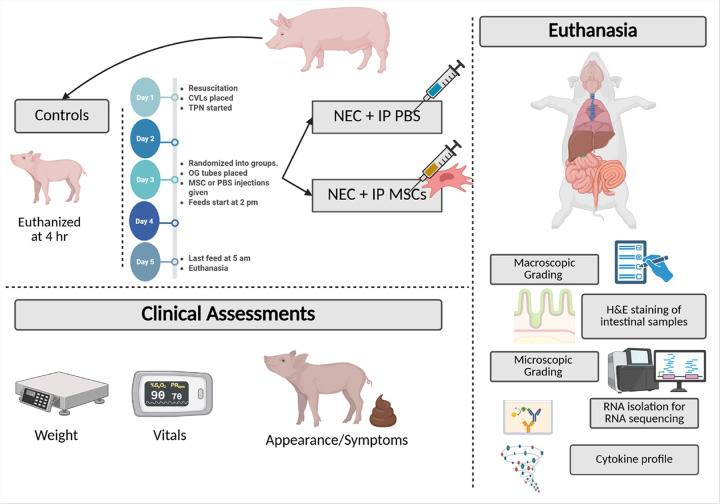
Purpose: Necrotizing enterocolitis (NEC) is a life-threatening gastrointestinal disease of prematurity characterized by inflammation, necrosis, and high morbidity. Current therapies are limited, necessitating the development of novel treatments. Mesenchymal stromal cells (MSCs) have shown promise in murine NEC models. Given the anatomical and physiological similarities between premature piglets and human infants, we employed a preterm piglet model to evaluate MSC efficacy. We hypothesized that intraperitoneal MSC administration would reduce intestinal injury in NEC.

Methods: Preterm piglets were delivered via cesarean section. NEC was induced on day 3 through hypertonic enteral feeding. MSCs were administered intraperitoneally at low, medium, or high doses. Piglets were monitored and euthanized based on clinical criteria. Clinical scores, weight change, gross and histologic intestinal injuries were assessed. Cytokine levels in serum and ileum were quantified via ELISA, and intestinal tissue was analyzed by RNA sequencing. Statistical significance was set at p < 0.05.

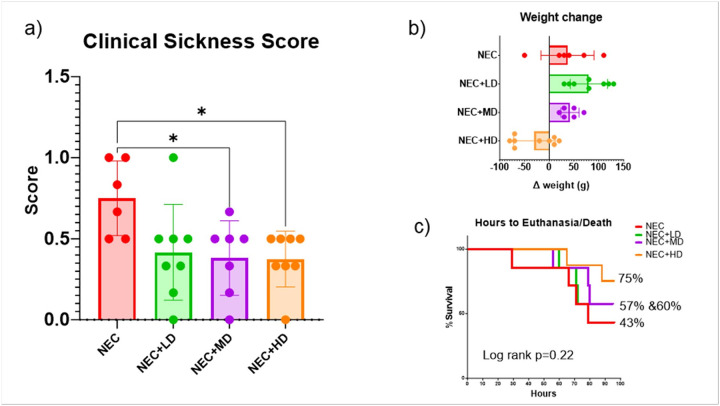
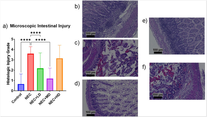
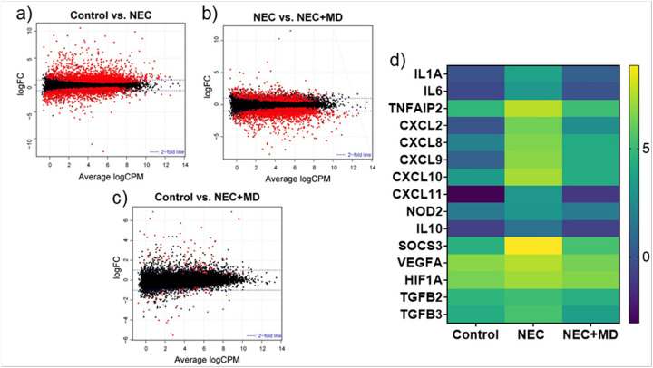
Results: Medium-dose MSCs significantly improved clinical scores and reduced both gross and histologic intestinal injury (p < 0.05). A corresponding decrease in pro-inflammatory cytokines was observed.

Conclusion: This is the first study to demonstrate therapeutic benefit of MSCs in a preterm piglet NEC model, supporting their potential use in translational NEC therapies.

Keywords: Inflammation; Mesenchymal Stromal Cells; Necrotizing Enterocolitis; Preterm Piglet; Stem Cell Therapy.


Conflict of interest statement

Conflicts of Interest / Competing Interests The authors declare no competing interests.

Figures

Figure 1
Figure 1
The Premature Piglet NEC Model. Schematic representation of the preterm piglet model of necrotizing enterocolitis (NEC) and treatment with intraperitoneal (IP) mesenchymal stromal cells (MSCs). Control piglets received resuscitation but were euthanized 4 hours after delivery without enteral feeding. Experimental piglets received central venous lines (CVLs) and total parenteral nutrition (TPN) on Day 1, were randomized to receive IP injections of either phosphate-buffered saline (PBS) or human MSCs on Day 3, and began enteral feeding on Day 3. Clinical assessments, including weight, vitals, and appearance/symptoms, were recorded daily. On Day 5, after the final feed, piglets were euthanized for sample collection. Post-mortem evaluations included gross and microscopic intestinal grading, hematoxylin and eosin (H&E) staining, cytokine profiling, and RNA sequencing of intestinal tissue. Created in https://BioRender.com
Figure 2
Figure 2
Clinical Sickness Score, Weight Gain/Loss, Survival. (a) Clinical sickness scores were significantly reduced in animals treated with mid-dose (NEC+MD) and high-dose (NEC+HD) MSCs compared to NEC animals (p=0.0447 and p=0.00335, respectively). NEC+LD animals also trended toward improvement. Asterisks indicate statistically significant differences. (b) Weight change from birth to euthanasia is shown for each group. MSC-treated animals showed less weight loss compared to untreated NEC controls, with the greatest preservation of weight observed in the NEC+HD group. (c) Kaplan–Meier survival analysis showing percentage survival over time for NEC animals with or without MSC treatment. Although a higher proportion of animals survived in the NEC+HD group (75%) compared to NEC (43%), the differences did not reach statistical significance (Log-rank p = 0.22). Final survival percentages are listed beside each group
Figure 3
Figure 3
Macroscopic Intestinal Injury. (a) Macroscopic intestinal injury scores were significantly elevated in NEC animals compared to controls (p <0.0001). Treatment with mid-dose MSCs (NEC+MD) significantly reduced injury scores compared to NEC animals (p = 0.0431), while low-dose (NEC+LD) and high-dose (NEC+HD) groups showed similar trends but did not reach statistical significance. (b-f) Representative gross intestinal images from each experimental group. (b) Control animal with normal bowel appearance and meconium in colon. (c) NEC animal with severe distension, necrosis, and discoloration. (d) NEC+LD with moderate edema and focal discoloration. (e) NEC+MD with mild edema and preserved coloration. (f) NEC+HD with moderate distension and severe congestion and inflammation
Figure 4
Figure 4
Microscopic Intestinal Injury. (a) Microscopic intestinal injury grades were significantly increased in NEC animals compared to controls (p = <0.0001). Treatment with low- and mid-dose MSCs significantly reduced histologic injury compared to NEC animals (both p<0.0001). No significant difference was observed in the NEC+HD group. (b-f) Representative hematoxylin and eosin (H&E) stained sections at 10× magnification (scale bars = 100 μm). (b) Control tissue with normal villous architecture and no inflammation. (c) NEC tissue showing marked epithelial sloughing, inflammation, and villous blunting. (d) NEC+LD showing partial preservation of architecture with reduced inflammation. (e) NEC+MD demonstrating near-normal mucosal structure. (f) NEC+HD with mild to moderate mucosal damage
Figure 5
Figure 5
Serum and Intestinal Cytokine Profile. Pro- inflammatory cytokine levels were measured in serum (top row) and terminal ileum tissue (bottom row) from control, NEC, and NEC+MSC mid-dose (NEC+MD) animals. Serum values are shown in pg/mL, and tissue levels are normalized to total protein (pg/ng). Asterisk on graph indicated significant relationships. Significant relationships: Top row – Serum cytokines: IL-6: NEC vs. Control (p = 0.0026), NEC+MD vs. NEC (p = 0.0286); IL-17A: NEC+MD vs. NEC (p=0.0271); TNF-α: NEC vs. Control (p = 0.0338). Bottom row – Terminal ileum cytokines: IL-6: NEC vs. Control (p <0.0001), NEC+MD vs. NEC (p<0.0001); IL-17A: NEC vs. Control (p = 0.0047), NEC+MD vs. NEC (p = 0.0084); TNF-α: NEC+MD vs. NEC (p = 0.0064)
Figure 6
Figure 6
Transcriptomic Analysis of Piglet Intestinal Tissues. (a–c) Smear plots generated from RNA sequencing data showing differentially expressed genes (DEGs) across experimental groups. Each point represents an individual gene; red dots indicate genes with a false discovery rate (FDR) < 0.05. (a) Control vs. NEC. (b) NEC vs. NEC+MD. (c) Control vs. NEC+MD. (d) Heatmap of selected inflammation- and injury-associated genes. Expression values are log-transformed, normalized counts. Genes were selected based on having significantly increased expression in NEC and reduction following MSC treatment. Cytokines: IL1A, IL6, and IL10. Chemokines: CXCL2, CXCL8 (also known as IL-8), CXCL9, CXCL10, and CXCL11. Immune signaling regulators: TNFAIP2, SOCS3, and NOD2. Hypoxia and growth factor-related genes: VEGFA, HIF1A, TGFB2, and TGFB3

Similar articles

References

    1. Fitzgibbons SC et al. (2009) Mortality of necrotizing enterocolitis expressed by birth weight categories. J Pediatr Surg, 44(6): pp. 1072–5; discussion 1075–6 - PubMed
    1. Moschino L et al. (2021) Optimizing Nutritional Strategies to Prevent Necrotizing Enterocolitis and Growth Failure after Bowel Resection. Nutrients, 13(2) - PMC - PubMed
    1. Meister AL, Doheny KK, Travagli RA (2020) Necrotizing enterocolitis: It’s not all in the gut. Exp Biol Med (Maywood) 245(2):85–95 - PMC - PubMed
    1. Hunter CJ et al. (2008) Understanding the susceptibility of the premature infant to necrotizing enterocolitis (NEC). Pediatr Res 63(2):117–123 - PubMed
    1. Neu J, Walker WA (2011) Necrotizing enterocolitis. N Engl J Med 364(3):255–264 - PMC - PubMed

Publication types

LinkOut - more resources