[Preprint]. 2025 Aug 20:rs.3.rs-5470731.
doi: 10.21203/rs.3.rs-5470731/v1.

Paclitaxel neurotoxicity is triggered by epidermal EG5-dependent microtubule fasciculation and X-ROS formation

Affiliations

Paclitaxel neurotoxicity is triggered by epidermal EG5-dependent microtubule fasciculation and X-ROS formation

Chia-Jung Hsieh et al. Res Sq. .

Abstract

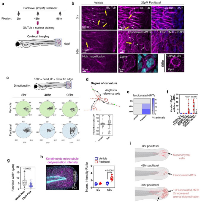
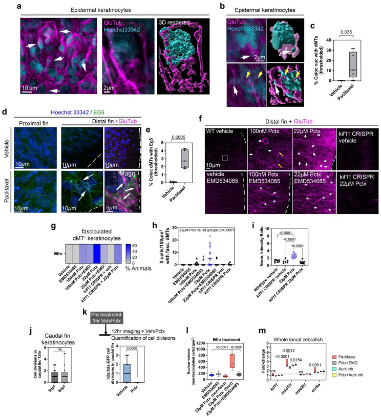
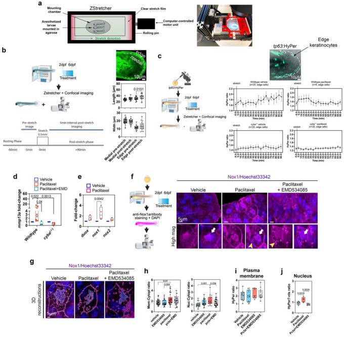
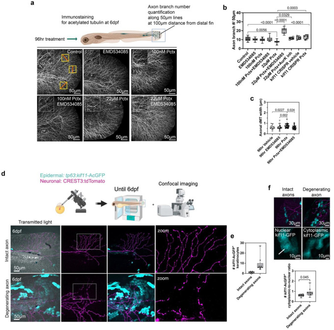
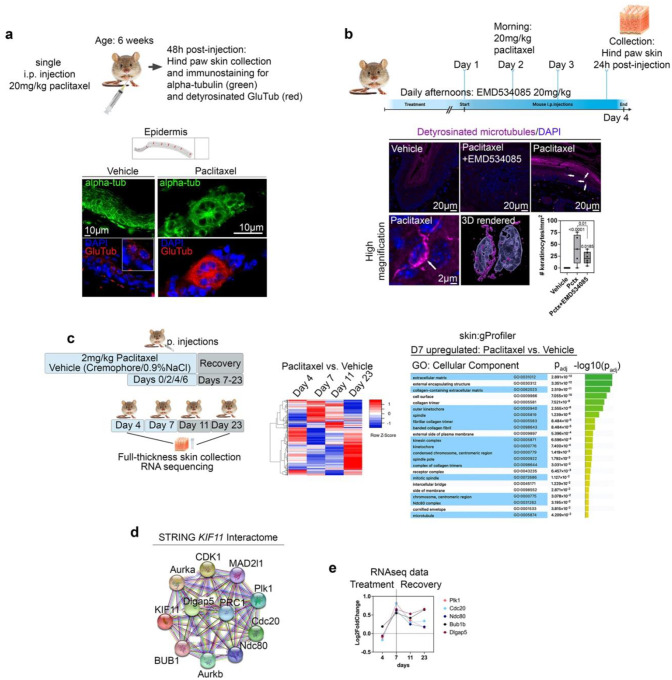
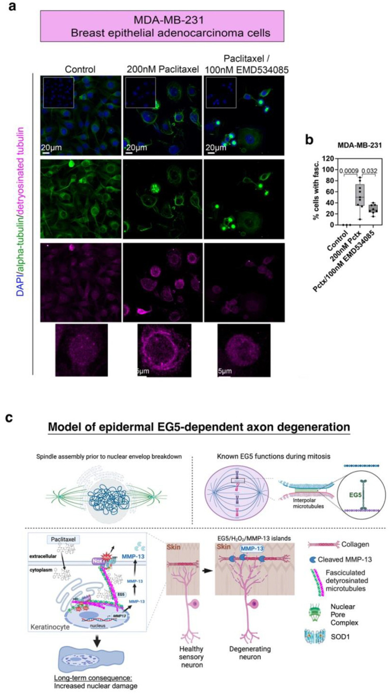
Taxanes are frontline chemotherapeutics that stabilize microtubules, induce mitotic arrest, and drive tumor remission. However, their off-target effects in healthy tissues, most notably cutaneous axon degeneration underlying chemotherapy-induced peripheral neuropathy (CIPN), remain poorly understood. Here, we show that paclitaxel induces microtubule fasciculation in epidermal keratinocytes through the mitotic kinesin Eg5, thereby initiating CIPN. Mechanistically, paclitaxel enhances Eg5-dependent fasciculation of detyrosinated (stabilized) microtubules, which constrict and breach the nuclear lamina. This deformation triggers tension-dependent NADPH oxidase-mediated nuclear ROS (X-ROS) formation upstream of mmp13 transcription, a pathway we previously demonstrated drives sensory axon degeneration. Employing a cross-species framework spanning zebrafish, mice, human skin biopsies, and a breast adenocarcinoma cell line, we uncover a conserved paclitaxel-Eg5 mechanism leading to fasciculation of stable microtubules in both healthy epidermis and cancer cells. These findings highlight the dualistic nature of paclitaxel action and underscore the challenge of preserving anticancer efficacy while preventing neurotoxic side effects.

Keywords: C57BL; Eg5; Kif11; Kinesin-5; MMP13; Taxol; cell cycle; detyrosination; fasciculation; keratinocyte; mice; microtubules; nucleus; paclitaxel; patient; peripheral neuropathy; zebrafish.


Conflict of interest statement

Competing interests. The authors have no competing interests in the research.

Figures

Figure 1.
Figure 1.. Detyrosination and keratinocyte-specific microtubule fasciculation following paclitaxel treatment.
(a) Treatment timeline and dMT detection strategy. (b) Weak axonal detyrosination (yellow arrows) and strong MSc labeling (asterisks) are observed in both 3hr vehicle- and paclitaxel-treated fish. Axonal detyrosination becomes more pronounced after 96hr paclitaxel treatment (yellow arrows), and keratinocytes display dMT fasciculation (white arrows; see Movies S2, S3). (c) Polar plots depicting the orientation and curvature of fasciculated dMTs in individual keratinocytes (180°=proximal; 0° = distal fin) show increased curvature in keratinocytes from 96hr paclitaxel-treated animals (n=4 animals/plot). (d) Angular deviation from a reference axis (Imaris), with 0° indicating straight alignment. (e) Increased percentage of animals with fasciculated dMTs following 48hr and 96hr paclitaxel treatment, but not at 3hr or in vehicle controls (n≥15 animals/group). (f) Paclitaxel (22μM) for 48hr or 96hr significantly increases the number of caudal fin keratinocytes with fasciculated dMTs compared to vehicle and 100nM paclitaxel (n≥17 animals/group). (g) 96hr treatment with 100nM paclitaxel results in wider dMT fascicles compared to 22μM paclitaxel (n=5 animals/group). (h) Normalized intensity ratios of detyrosinated microtubules in keratinocytes quantified in the indicated ROI (arrowhead) of the caudal fin. GluTub signal was normalized to Hoechst 33342 nuclear staining within each fin. (i) Schematic summary of observations.
Figure 2.
Figure 2.. Fasciculation of dMTs is EG5 dependent.
(a) Low- and high-magnification images and 3D rendering of fasciculated dMTs (anti-GluTub) and their association with keratinocyte nuclei (Hoechst33342) (arrows). (b) Left: Fluorescence images of Hoechst/detyrosinated MTs. Right: 3D rendering using Imaris surface rendering. Fasciculated dMTs pinch off nuclear content (white arrows, top panel) or pierce the nucleus (bottom) (Movies S5,6). (c) Significant colocalization of Hoechst33342 nuclei with GluTub fluorescence (thresholded) in keratinocytes of paclitaxel-treated versus vehicle-treated fish (n=6 animals/group). (d) EG5 staining shows puncta in proximal caudal fin keratinocytes of both vehicle- and paclitaxel-treated animals. Dashed lines indicate the distal fin margin. Paclitaxel treatment promotes EG5 localization in aster-like structures (arrows) and co-localization with fasciculated dMTs (white arrows, magnified image). (e) EG5-GluTub colocalization is significantly increased following paclitaxel treatment compared with vehicle. (f) Co-administration of EMD534085 with 100nM or 22μM paclitaxel prevents microtubule fasciculation observed with paclitaxel alone (asterisks: detyrosinated MScs; yellow arrows: axons; white arrows: fasciculated dMTs). (g) Fasciculated dMTs are observed in paclitaxel-treated animals but not in those co-treated with EMD534085 or in kif11 CRISPR knockouts (n=23 animals/group). (h) Number of keratinocytes per animal with fasciculated dMTs is reduced with EMD534085 co-administration (n=21 animals/group). (i) Normalized GluTub intensity ratios in vehicle- and paclitaxel-treated wild-type and kif11 CRISPR knockout animals (n=21 animals/group). (j) Keratinocyte divisions at 3dpf (n=28) and 6dpf (n=12) assessed via time-lapse imaging in Tg(h2a:h2a-GFP) fish. (k) Keratinocyte divisions over 12hr at 3dpf with vehicle or paclitaxel treatment (n=9 animals/group). (l) Nuclear volume increases after 96hr paclitaxel treatment and is rescued by EMD534085 co-treatment. (m) qPCR of whole larvae after 96hr treatment shows significant mad1l1 and aurka upregulation, with mad1l1 reduced by EMD534085 co-treatment (n=10 animals/group, 3 biological replicates).
Figure 3.
Figure 3.. Eg5-dependent nuclear X-ROS formation upstream of mmp13 transcription.
(a) ZStretcher design for simultaneous stretching and confocal imaging of live zebrafish. (b) Outer fin edge keratinocytes in Tg(tp63:AcGFP-CAAX) fish show increased length but not width following stretch, while medially located keratinocytes are unaffected (separation by dotted line). (c) Ratiometric HyPer imaging from pre-stretch to 90min post-stretch in 5-min intervals in wildtype (top panel) and cyba−/− mutants (bottom panel), treated with either vehicle (left) or paclitaxel (right), demonstrates faster HyPer oxidation in paclitaxel-treated wildtype animals compared with respective vehicle controls. HyPer oxidation is largely absent or reduced in paclitaxel-treated cyba−/− mutants relative to vehicle and wildtype controls (n=5–9 animals/treatment). (d) qPCR demonstrates enhanced mmp13 expression in wildtype fish treated with paclitaxel, but not in cyba−/− mutants (n=10 animals/group in 3 biological replicates). EMD534085 co-administration with paclitaxel in wildtype fish prevents mmp13a upregulation. (e) qPCR shows significantly increased nox1 expression following 48hr paclitaxel treatment (10 animals/group and 3 biological replicates). (f) Nox1 expression in the caudal fin of a 96hr vehicle-treated control animal shows Nox1 localization along plasma membranes. Additional nuclear Nox1 recruitment following 96hr paclitaxel treatment is abolished when EMD534085 is co-administered. (g) 3D reconstruction of Nox1 staining shown in (f) (Movies 9, 10). (h) Nox1 membrane:cytoplasmic and nuclear:cytoplasmic ratios are significantly increased following 96hr paclitaxel treatment and reduced with EMD534085 co-administration (n=6–9 animals/group). (i) HyPer oxidation measured at the plasma membrane does not differ among treatment groups. (j) HyPer7-nls oxidation is significantly increased with paclitaxel compared with EMD534085 co-administration and vehicle (n=5 animals/group).
Fig. 4.
Fig. 4.. Keratinocyte-specific EG5 activity induces paclitaxel neurotoxicity.
(a) Immunostaining for acetylated tubulin reveals cutaneous sensory axon degeneration with 22μM (but not 100nM) paclitaxel, and rescue when EMD534085 is co-administered (n≥11 animals/group). (b) Quantification of axon branch number in caudal fin along lines (marked in red with yellow box) shows that both AurK inhibitor and kif11 CRISPR knockout rescue paclitaxel-induced neurotoxicity similarly to EMD534085. Notably, EMD534085 also promotes axon regrowth (n=6 animals/group). (c) dMT width in axons is significantly increased following 22 μM paclitaxel treatment and is rescued by EMD534085 co-administration (n=4–5 animals/group). (d) Top: Expression of isl1:LexA-lexAop_14xUAS-tdTomato (magenta) in cutaneous sensory neurons shows intact axons when tp63:kif11-AcGFP (blue) keratinocytes are absent. Bottom: Axon degeneration is apparent when tp63:kif11-AcGFP-expressing basal keratinocytes are adjacent to isl1:tdTomato-positive axons. (e) The degree of axon degeneration correlates with the number of keratinocytes expressing kif11:GFP (n=14 animals). (f) Axon degeneration in the caudal fin correlates with increased cytoplasmic EG5-GFP localization (n=14 animals). Nuclear Eg5-AcGFP is marked with an arrow.
Figure 5.
Figure 5.. Paclitaxel/Eg5-dependent dMT fasciculation and cell cycle gene expression changes in mouse skin.
(a) Immunostaining for alpha-tubulin and GluTub in the epidermis of 6-week-old mice following a single vehicle or paclitaxel injection. Fasciculated dMTs and multinucleated rosettes are apparent in paclitaxel-injected epidermis. (b) Administration of paclitaxel+EMD534085 significantly reduces dMT fasciculation induced by paclitaxel alone. (c) RNAseq of hind paw skin from mice injected with vehicle or paclitaxel reveals expression changes across different days, prior to pain (D4), peak pain (D7,11), and recovery (D23). Functional enrichment analysis at D7 identifies significant GO term enrichment in extracellular matrix, collagen, and cell cycle processes. (d) STRING interactome analysis of mitosis-related genes from our RNAseq dataset highlights interactions with KIF11. (e) Co-upregulation of cell cycle checkpoint regulators during peak pain (D7/11) identified through STRING (d).
Figure 6.
Figure 6.. Breast cancer cells show conserved features to zebrafish and mouse keratinocytes.
(a) MDA-MB-231 breast cancer cells stained for alpha-tubulin and GluTub show increased fasciculation of dMTs following paclitaxel treatment. This phenotype is rescued when EMD534085 is co-administered. (b) The percentage of cells per field of view (identical 1024×1024 pixels) with dMT fasciculation is significantly increased upon treatment with 200nM paclitaxel for 24hr compared with vehicle and paclitaxel+EMD534085 treatment. (c) Model for EG5-dependent paclitaxel neurotoxicity. Top: Spindle assembly occurs during prophase prior to nuclear envelope breakdown. Eg5 may have a similar crosslinking function when activated by paclitaxel. Bottom: Paclitaxel promotes detyrosination (long-term stabilization) of microtubules in prophase keratinocytes, inducing EG5-dependent fasciculation near the nucleus. This creates microtubule tension, activating Nox1-dependent X-ROS (H2O2) formation. X-ROS are secreted into the nucleus via nuclear pores or envelope disruptions, triggering MMP13 transcription, which promotes MMP-13-dependent ECM degradation and axon degeneration

Similar articles

References

    1. Gornstein E. & Schwarz T. L. The paradox of paclitaxel neurotoxicity: Mechanisms and unanswered questions. Neuropharmacology 76 Pt A, 175–183 (2014). 10.1016/j.neuropharm.2013.08.016 - DOI - PubMed
    1. Argyriou A. A., Bruna J., Genazzani A. A. & Cavaletti G. Chemotherapy-induced peripheral neurotoxicity: management informed by pharmacogenetics. Nat Rev Neurol 13, 492–504 (2017). 10.1038/nrneurol.2017.88 - DOI - PubMed
    1. Argyriou A. A., Bruna J., Marmiroli P. & Cavaletti G. Chemotherapy-induced peripheral neurotoxicity (CIPN): an update. Crit Rev Oncol Hematol 82, 51–77 (2012). 10.1016/j.critrevonc.2011.04.012 - DOI - PubMed
    1. Argyriou A. A. et al. Neurophysiological, nerve imaging and other techniques to assess chemotherapy-induced peripheral neurotoxicity in the clinical and research settings. Journal of neurology, neurosurgery, and psychiatry (2019). 10.1136/jnnp-2019-320969 - DOI - PubMed
    1. Argyriou A. A., Zolota V., Kyriakopoulou O. & Kalofonos H. P. Toxic peripheral neuropathy associated with commonly used chemotherapeutic agents. J BUON 15, 435–446 (2010). - PubMed

Publication types

LinkOut - more resources