

This is a preprint.
A direct method for imaging gradient levels of retinal hypoxia in a model of retinopathy of prematurity (ROP)
- PMID: 40894049
- PMCID: PMC12393608
- DOI: 10.21203/rs.3.rs-7247191/v1
A direct method for imaging gradient levels of retinal hypoxia in a model of retinopathy of prematurity (ROP)
Update in
-
A direct method for imaging gradient levels of retinal hypoxia in a model of retinopathy of prematurity (ROP).BMC Ophthalmol. 2026 Jan 6;26(1):61. doi: 10.1186/s12886-025-04601-y. BMC Ophthalmol. 2026. PMID: 41495706 Free PMC article.
Abstract
Background: Retinal hypoxia may contribute to the development of preretinal neovascularization in patients with retinopathy of prematurity (ROP). Ciliary bodies compensate oxygen delivery to the retina, and the levels of hypoxia may vary across the peripheral avascular area in ROP. In this study, we have investigated a direct method for imaging gradient levels of retinal hypoxia at the peripheral avascular retina using a model ROP.
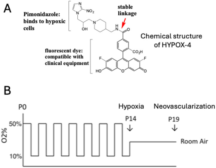
Methods: The rat 50/10 oxygen-induced retinopathy (OIR) model was generated by exposing the newly born Brown-Norway rat pups to a 24 hours alternate cycles of 50% and 10% oxygen for 14 days. We also confirmed the development of neovascularization in this model. HYPOX4 was used as a direct method for imaging gradient levels of retinal hypoxia at the peripheral avascular retina. A separate group of rat OIR pups were used to confirm gradient levels of retinal hypoxia using pimonidazole immunostaining. Gradient levels of retinal hypoxia was analyzed using ImageJ software from fluorescence intensities of HYPOX-4 and Pimonidazole immunostaining.
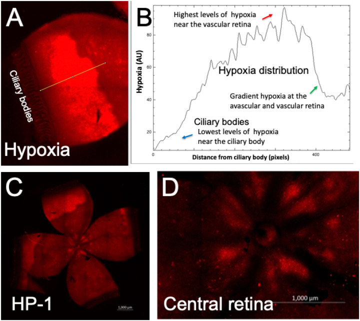
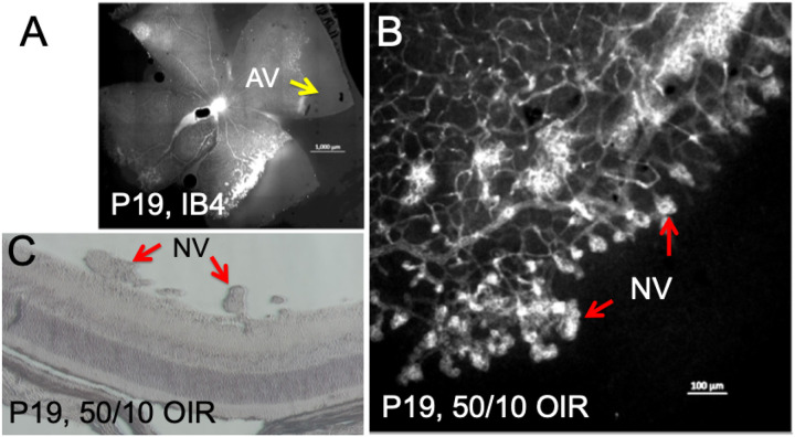
Results: Retinal hypoxia was observed in the peripheral avascular retinas in rat OIR. Based on fluorescence intensity measurements, retinal hypoxia was at minimal levels near the ciliary bodies. Retinal hypoxia was at its maximum levels towards the avascular-vascular transition zones. Interestingly, we observed hemiretinal avascular retina temporal to the optic nerve in this OIR model, similar to human ROP retinas. In the retinal cross-section, hypoxia was not detectable near the ora serrata in rat OIR may be due to oxygen delivery by the ciliary bodies. Both pimonidazole and HYPOX-4 showed similar patterns of retinal hypoxia at the peripheral avascular retina in this model. As expected, preretinal neovascularization was observed at the avascular-vascular transition zones arising from the existing retinal vascular structures in this OIR model in Brown-Norway rats.
Conclusions: In this study, we have characterized gradient levels of retinal hypoxia in the rat model of 50/10 OIR using a direct method from HYPOX-4 fluorescence. We observed minimal levels of retinal hypoxia near the ciliary bodies in this model and increased towards the avascular-vascular transition zones. In addition, we observed that the central vascularized retina remains gradient hypoxic in this model which could be detected using HYPOX-4. This study may clarify our understanding of retinal hypoxia in the ROP patient at the peripheral retinas.
Keywords: HYPOX-4; ROP; Retinopathy of prematurity; fluorescence imaging; molecular imaging; optical imaging; retinal hypoxia.
Conflict of interest statement
Competing interests: The authors declare that they have no conflicts of interest to report.
Figures


References
-
- Fevereiro-Martins M, Marques-Neves C, Guimaraes H, Bicho M. Retinopathy of prematurity: A review of pathophysiology and signaling pathways. Surv Ophthalmol. 2023;68(2):175–210. - PubMed
-
- Selvam S, Kumar T, Fruttiger M. Retinal vasculature development in health and disease. Prog Retin Eye Res. 2018;63:1–19. - PubMed
Publication types
Grants and funding
LinkOut - more resources
Full Text Sources
Research Materials
