. 2025 Jul 21:12:1520101.
doi: 10.3389/fmolb.2025.1520101. eCollection 2025.

RNA sequencing-based profiling of differentially expressed microRNAs in endothelial cells from offspring of hypertensive pregnancies: a preliminary study

Affiliations

RNA sequencing-based profiling of differentially expressed microRNAs in endothelial cells from offspring of hypertensive pregnancies: a preliminary study

Nurul Iffah Mohd Isa et al. Front Mol Biosci. .

Abstract

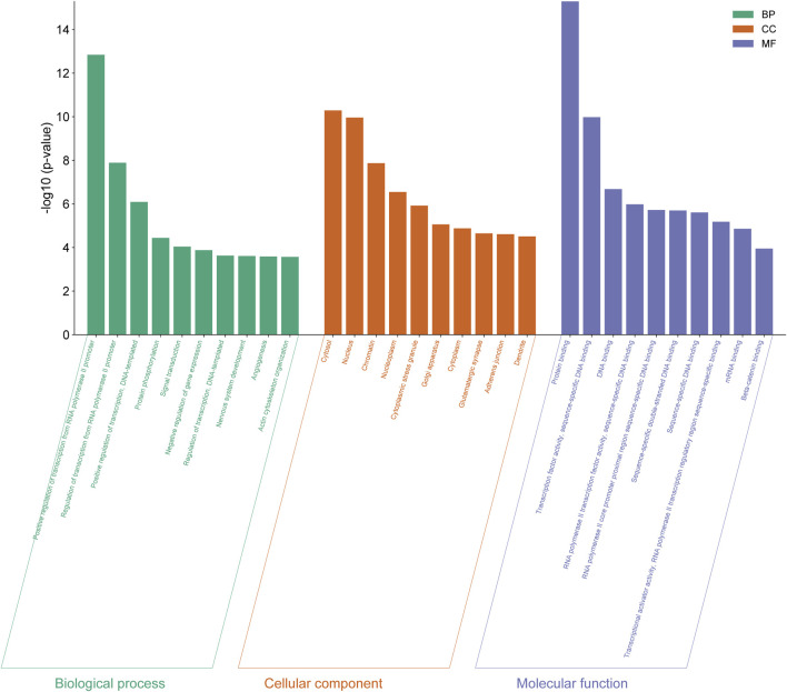
Offspring of mothers with hypertensive disorders of pregnancy (HDP) are at increased risk of developing endothelial dysfunction and cardiovascular disease (CVD) in adulthood. MicroRNAs (miRNAs), as key regulators of endothelial cells, may contribute to the early onset of endothelial dysfunction. However, there are limited studies characterizing the miRNA profile of endothelial cells in offspring of HDP. Therefore, this study aims to determine the miRNA expression profile of human umbilical vein endothelial cells (HUVECs) isolated from the offspring of HDP. HUVECs were obtained from both normal and hypertensive umbilical cords. RNA sequencing analysis revealed that eight miRNAs were significantly upregulated in HUVECs from HDP (p < 0.05). The target genes of these miRNAs were then predicted using four databases: miRDB, TargetScan, DIANA-microT-CDS, and miRWalk. Gene ontology, pathway enrichment, and protein-protein interaction network analyses revealed that the target genes of these miRNAs are involved in cellular functions and pathways related to angiogenesis and cellular senescence, which may contribute to endothelial dysfunction and CVD. The most significantly upregulated miRNA, hsa-miR-196a-5p expression was then validated through stem-loop RT-qPCR where its expression was significantly upregulated in hypertensive HUVEC by 6-fold as compared to normal HUVEC (p < 0.01). These findings offer insights into the role of miRNAs in the development of CVD in offspring exposed to HDP, highlighting their potential as predictive markers and therapeutic targets in the future.

Keywords: RNA sequencing; cardiovascular disease; endothelial dysfunction; human umbilical vein endothelial cells; hypertensive disorders of pregnancy; microRNA.


Conflict of interest statement

The authors declare that the research was conducted in the absence of any commercial or financial relationships that could be construed as a potential conflict of interest.

Figures

FIGURE 1
FIGURE 1
Expression of vWF in HUVEC isolated from umbilical cord via immunofluorescence staining. (A) Cobblestone morphology of the cells was seen under the inverted microscopy (×100 magnification) (B) Positive expression of vWF in HUVECs was detected and the nucleus of the cells was stained with DAPI (×400 magnification). The scale bar of the pictures was set at 100 µm.
FIGURE 2
FIGURE 2
Venn diagrams of (A) known and (B) unique miRNAs identified in normal and PIH-HUVECs. (C) A volcano plot showing the miRNA expression profile in HUVECs, with the red dots representing significantly upregulated miRNAs. miRNAs that do not show significant expression changes are shown as blue dots. (D) Hierarchical clustering and heatmaps of miRNA expression profiles. The samples are arranged vertically and clustered using colour bars between the dendrogram and the heatmap. The colour key represents the relative expression of miRNAs in all samples. Red stands for higher expression levels, blue for lower expression levels.
FIGURE 3
FIGURE 3
Determination of target gene numbers illustrated by Venn diagram which obtained from four databases including DIANA-microT-CDS, TargetScan, miRDB, and miRWalk for all differentially expressed miRNAs.
FIGURE 4
FIGURE 4
GO functional analysis of differentially expressed genes from eight differentially expressed miRNAs in normal vs PIH groups where GO were classified into three categories including biological process (BP), cellular components (CC), and molecular function (MF).
FIGURE 5
FIGURE 5
Scatter plot showing the differentially expressed genes by KEGG pathway analysis. The size of the dot represents the number of genes involved in the pathway and the range of the p-value is determined by the colour of the dot. The degree of enrichment is determined by the fold enrichment and the significance of the enrichment is determined by the p-value. A higher level of enrichment indicates a higher degree of enrichment, while a lower p-value indicates a higher significance of enrichment.
FIGURE 6
FIGURE 6
PPI network between the top 20 target genes expressed by the differentially expressed miRNAs illustrated via CytoHubba in CytoScape. Colour intensity of the nodes from red to yellow indicates for ranking of the nodes by degree ranking method (red to yellow–high to low).
FIGURE 7
FIGURE 7
Interaction network of hub genes with the differentially expressed miRNAs, biological process (BP) in GO and pathways using ClueGO. The colour of the nodes represents for the cluster of GO and pathway. Triangle node represents for miRNA, ellipse node represents for the genes, square node represents for pathway, and hexagon node represents for BP in GO. All of the pathways and functions depicted in the figure are statistically significant (p < 0.05).
FIGURE 8
FIGURE 8
Validation of miR-196a-5p expression in normal vs PIH HUVEC via stem-loop RT-qPCR. Values are shown as mean ± SEM, n = 6, **p < 0.01 vs normal. Result was analyzed using Independent T-test.

References

    1. Abdull Sukor A. N., Ankasha S. J., Ugusman A., Aminuddin A., Mokhtar N. M., Abidin Z., et al. (2022). Impact of offspring endothelial function from de novo hypertensive disorders during pregnancy: an evidence-based review. Front. Surg., 9. 967785. 10.3389/fsurg.2022.967785 - DOI - PMC - PubMed
    1. Alsnes I. V., Vatten L. J., Fraser A., Bjørngaard J. H., Rich-Edwards J., Romundstad P. R., et al. (2017). Hypertension in pregnancy and offspring cardiovascular risk in young adulthood: prospective and sibling studies in the HUNT study (Nord-Trøndelag Health Study) in Norway. Hypertension 69 (4), 591–598. 10.1161/hypertensionaha.116.08414 - DOI - PubMed
    1. Arderiu G., Espinosa S., Peña E., Aledo R., Badimon L. (2015). PAR2–SMAD3 in microvascular endothelial cells is indispensable for vascular stability via tissue factor signaling. J. Mol. Cell Biol. 8 (3), 255–270. 10.1093/jmcb/mjv065 - DOI - PubMed
    1. Bajpai D., Popa C., Verma P., Dumanski S. M., Shah S. (2023). Evaluation and management of hypertensive disorders of pregnancy. Kidney360 4, 1512–1525. 10.34067/kid.0000000000000228 - DOI - PMC - PubMed
    1. Barh D., Chaitankar V., Yiannakopoulou E. C., Salawu E. O., Chowbina S., Ghosh P., et al. (2014). In silico models. Anim. Biotechnol., 385–404. 10.1016/b978-0-12-416002-6.00021-3 - DOI

LinkOut - more resources