[Preprint]. 2025 Aug 18:2025.08.18.670552.
doi: 10.1101/2025.08.18.670552.

7 Tesla fMRI characterisation of the cortical-depth-dependent BOLD response in early human development

Affiliations

7 Tesla fMRI characterisation of the cortical-depth-dependent BOLD response in early human development

Jucha Willers Moore et al. bioRxiv. .

Abstract

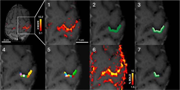
Human cortical development leading up to and around birth is crucial for lifelong brain function. Cortical activity can be studied using BOLD fMRI, however, previously limited sensitivity and spatial specificity has constrained understanding of how its emergence relates to functional cortical circuitry and neurovascular development at the mesoscale. To resolve this, we used ultra-high-field 7 Tesla MRI to acquire submillimetre resolution BOLD-fMRI data from 40 newborns and 4 adults. In all subjects, passive right-hand movement elicited localised, positive BOLD responses in contralateral primary somatosensory cortex. In newborns, depth-specific BOLD responses were still evident in the thinner cortex, with developmental changes in response temporal features and amplitudes at different depths. This provides insight into key rapidly evolving factors in early cortical development including neuronal function, vascular architecture, and neurovascular coupling. Our framework and findings provide a foundation for future studies of emerging cortical circuitry and how disruption leads to adverse outcomes.


Figures

Figure 1.
Figure 1.. Setup for fMRI data acquisition in neonates on the 7 T MRI system.
(A) Demonstration of neonate positioning inside the head coil at isocentre by foam cushions placed around the head. Infants are immobilised in a vacuum-evacuated blanket, and inflatable cushions were placed either side of the infant’s head to further prevent head motion. The MR-compatible pneumatically-driven wrist device which provides passive stimulation of the wrist inside the MRI scanner, fitted onto (B) a neonate wrist (C) and an adult wrist. (D) Exemplar fMRI acquisition field-of-view (pink) in neonates (top) and adults (bottom) overlaid onto a T2-weighted anatomical image. (E, F) Passive movement of the right wrist produces localised activation in the contralateral (left) primary somatosensory cortex in (E) one exemplar neonate (38 weeks PMA) and (F) adult (24 years). Exemplar activation maps showing significantly activated voxels (z > 3.1) following passive somatosensory stimulation of the right wrist (left). Mean BOLD timeseries (right) extracted from above-threshold voxels, with stimulation periods indicated in grey.
Figure 2.
Figure 2.. Region of interest (ROI) selection and cortical-depth bin definition for analysis of cortical-depth-dependent BOLD responses.
1. Significantly activated voxels in the contralateral primary somatosensory cortex following somatosensory stimulation of the right wrist. 2. A large ROI is manually defined (green) of the hand area of S1 including the most significantly activated voxels. The ROI spans the full thickness of the cortex across multiple EPI slices. 3. The ROI is split in into three “equivolume layers” or depth bins using LAYNII, where each shade of green indicates a depth bin, with light green being the most superficial depth bin. 4. Each slice is split into seven columns, denoted by colour. 5. The column and layer profiles are combined to produce columns with three equivolume cortical-depth bins, with colours corresponding to columns and shades corresponding to depth bins. 6. The activation map in units of BOLD percent signal change. 7. The columns in which the BOLD percent signal change in the deep cortical-depth bins was in the top 40th centile were selected to produce the final ROI used for the cortical-depth-dependent analyses, with depth bins denoted by shades, with the lightest shade representing the most superficial depth bin.
Figure 3.
Figure 3.. The cortical-depth-dependent BOLD response in S1 to passive somatosensory wrist stimulation is immature in the neonatal period and develops with age.
(Top) Exemplar significantly activated voxels (z>3.1) following sensorimotor stimulation to the right wrist and three equivolume cortical depths in the hand area of contralateral primary somatosensory cortex (S1) overlaid onto the mean EPI data. Lightest shade denotes the most superficial depth. Trial-average cortical depth dependent BOLD response timeseries (+/− SEM) in preterm, (n=8 neonates), early-term (n=8), and late-term (n=7) neonates, and adults (n=4).
Figure 4.
Figure 4.. Maturation of the BOLD response is cortical-depth specific.
(A) The cortical-depth profile of the maximum BOLD percent signal change across cortical depths averaged across all trials in preterm (n=8), early-term (n=8), and late-term (n=7) neonates, and adults (n=4). (B) Significantly different ratios of maximum BOLD precent signal change were found across age groups for Superficial:Middle (Kruskal-Wallis: H(3) = 35.12, p <0.001. Dunn post-hoc with Bonferroni adjustment: preterm vs. adult: p <0.001, early-term vs. adult: p <0.001, late-term vs. adult: p = 0.024) and superficial:Deep (Kruskal-Wallis: H(3) = 35.96, p < 0.001. Dunn post-hoc with Bonferroni adjustment: preterm vs. adult: p <0.001, early-term vs. late-term: p = 0.041). (* indicates p <0.05; ** indicates p <0.01; *** indicates p 0.001.)
Figure 5.
Figure 5.. Spatial extent of extravascular fields of large pial veins is not larger in the neonatal period compared with adults.
(A) The simulated field perturbation produced for a 65-μm diameter pial vein oriented perpendicular to B0 using the infinite-cylinder model of extravascular fields. A range of blood-tissue susceptibility (Δχblood-tissue) values (0.5, 1.0, 1.5 and 2.0 ppm) were simulated with 0.5 ppm being the adult-like value. A distance of 0 mm denotes the boundary between the vein and the grey matter tissue. (B) The predicted field perturbation for an adult-like large peripheral vein (orange; 65-μm diameter and 0.5 ppm Δχblood-tissue)and a neonate-like large peripheral vein (purple; 130-μm diameter and 1 ppm Δχblood-tissue).

References

    1. Allievi A. G. et al. Maturation of Sensori-Motor Functional Responses in the Preterm Brain. Cereb Cortex 26, 402–413 (2016). - PMC - PubMed
    1. Harris J. J., Reynell C. & Attwell D. The physiology of developmental changes in BOLD functional imaging signals. Dev Cogn Neurosci 1, 199–216 (2011). - PMC - PubMed
    1. Krsnik Ž., Majić V., Vasung L., Huang H. & Kostović I. Growth of Thalamocortical Fibers to the Somatosensory Cortex in the Human Fetal Brain. Frontiers in Neuroscience 11, 233 (2017). - PMC - PubMed
    1. Keunen K., Counsell S. J. & Benders M. J. N. L. The emergence of functional architecture during early brain development. NeuroImage 160, 2–14 (2017). - PubMed
    1. Kostović I. & Jovanov-Milošević N. The development of cerebral connections during the first 20–45 weeks’ gestation. Seminars in Fetal and Neonatal Medicine 11, 415–422 (2006). - PubMed

Publication types

LinkOut - more resources