[Preprint]. 2025 Aug 20:2025.08.19.671158.
doi: 10.1101/2025.08.19.671158.

Development of Degraders and 2-pyridinecarboxyaldehyde (2-PCA) as a recruitment Ligand for FBXO22

Affiliations

Development of Degraders and 2-pyridinecarboxyaldehyde (2-PCA) as a recruitment Ligand for FBXO22

Tian Qiu et al. bioRxiv. .

Abstract

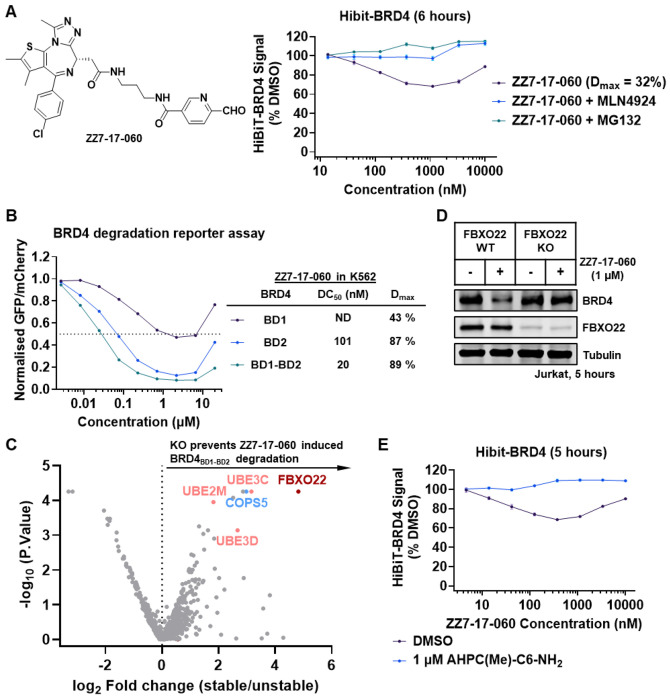
Targeted protein degradation (TPD) is a promising therapeutic strategy that requires the discovery of small molecules that induce proximity between E3 ubiquitin ligases and proteins of interest. FBXO22 is an E3 ligase that is overexpressed in many cancers and implicated in tumorigenesis. While FBXO22 was previously identified as capable of recognizing ligands bearing a primary amine degron, further investigation and development of recruitment ligands is required to enable its broader utility for TPD. Here, we describe the discovery of chemical probes that can either selectively degrade FBXO22 or recruit this ligase for TPD applications. First, we describe AHPC(Me)-C6-NH2 as a potent and selective FBXO22 degrader (DC50 = 77 nM, Dmax = 99%) that is suitable for interrogating the effects of FBXO22 loss of function. Further, we discovered that the simple hexane-1,6-diamine acts as a minimal FBXO22 self-degrader, whereas shorter C4 (putrescine) to C5 (cadaverine) analogs, found in mammalian cells, do not induce degradation. Finally, we found that 2-pyridinecarboxaldehyde (2-PCA) functions as a novel electrophilic degron capable of forming a reversible thioketal with cysteine 326 for recruiting FBXO22. Conjugating 2-PCA to various ligands successfully induced FBXO22-dependent degradation of BRD4 and CDK12. Collectively, these chemical probes will facilitate the study of FBXO22 biology and broaden its applicability in TPD.


Conflict of interest statement

Competing interests Nathanael S. Gray is a founder, science advisory board member (SAB) and equity holder in Syros, C4, Allorion, Lighthorse, Voronoi, Inception, Matchpoint, Shenandoah (board member), Larkspur (board member) and Soltego (board member). The Gray lab receives or has received research funding from Novartis, Takeda, Astellas, Taiho, Jansen, Kinogen, Arbella, Deerfield, Springworks, Interline and Sanofi. Benjamin L. Ebert has received research funding from Novartis and Calico. He has received consulting fees from Abbvie. He is a member of the SAB and shareholder for Neomorph Inc., Big Sur Bio, Skyhawk Therapeutics, and Exo Therapeutics. Eric S. Fischer is a founder, SAB member, and equity holder of Civetta Therapeutics, Proximity Therapeutics, Neomorph, Inc. (also board of directors), Stelexis Biosciences, Inc., Anvia Therapeutics, Inc. (also board of directors), CPD4, Inc (also board of directors) and Nias Bio, Inc. He is an equity holder in Avilar Therapeutics, Ajax Therapeutics (also SAB), Photys Therapeutics (also SAB), and Lighthorse Therapeutics. E.S.F. is a consultant to Novartis, EcoR1 capital and Deerfield. The Fischer lab receives or has received research funding from Deerfield, Novartis, Ajax, Interline, Bayer and Astellas. Katherine A. Donovan receives or has received consulting fees from Neomorph Inc and Kronos Bio.

Figures

Figure 1.
Figure 1.
Charting E3 ligand tethered with primary amine for FBXO22 degradation. A) Structure of ZXH-03-118. E3 ligase ligand is highlighted with yellow color, primary amine is highlighted with green color, spacer region is highlighted with blue color. B) General chemical structure of Thalidomide-Amide-Cn-NH2 and HiBiT-FBXO22 assay results for Jurkat cells treated with the CRBN ligand-based degraders for 5 hours. C) General chemical structure of AHPC(R)-Cn-NH2 and HiBiT-FBXO22 assay results for Jurkat cells treated with the VHL ligand-based degraders for 5 hours.
Figure 2.
Figure 2.
(S,R,S)-AHPC(Me)-C6-NH2 is a potent and selective FBXO22 degrader. A) Chemical structure of AHPC(Me)-C6-NH2. B) Western blots showing FBXO22 degradation in Jurkat cells treated with the indicated concentration of AHPC(Me)-C6-NH2 for 5 hours. C) Quantification of the data shown in B. D) Western blots showing FBXO22 degradation in Jurkat cells treated with 1 μM AHPC(Me)-C6-NH2 at indicated time. E) Quantification of the data shown in D. F) Quantitative proteome-wide mass spectrometry in MOLT-4 cells after 5 hours treatment with 1 μM AHPC(Me)-C6-NH2.
Figure 3.
Figure 3.
Mechanism of AHPC(Me)-C6-NH2 induced FBXO22 degradation. A) Chemical structures of ZZ7-23-033, ZZ7-23-034 and AHPC-CHO. B) Western blots showing FBXO22 degradation in Jurkat cells treated with the indicated compounds for 5 hours. C) Western blots showing FBXO22 degradation in Jurkat cells pre-treated with the indicated inhibitors for 1 hour, followed by treatment with 1 μM AHPC(Me)-C6-NH2 for 5 hours. D) TR-FRET assay to measure molecule-dependent ternary complex formation between VCB complex and SKP1/FBXO22 (WT or C326A). Each point represents a duplicate replicates; the mean value line is drawn. E) Scatterplots depicting relative protein abundance following Flag-VHL-EloB/C enrichment from in-lysate treatment with 1 μM AHPC-CHO and recombinant Flag-VHL-EloB/C spike in. Scatterplots display fold change in abundance to DMSO. Significant changes were assessed by moderated t-test as implemented in the limma package with log2 FC shown on the y-axis and log10 (P.value) on the x-axis. F) Western blots showing protein levels of FBXO22 and its downstream signals in HEK293T and THP-1 cells treated with indicated concentration of AHPC(Me)-C6-NH2 and AHPC-CHO for 24 hours.
Figure 4.
Figure 4.
Primary diamine is FBXO22 self-degrader. A) Schematic illustration showing the concept of FBXO22 self-degrader. B) Metabolic conversion of primary diamine to dialdehyde. C) HiBiT-FBXO22 assay results for Jurkat cells treated with the primary diamines for 5 hours. D) Western blots showing FBXO22 degradation in Jurkat cells pre-treated with the indicated inhibitors for 1 hour, followed by treatment with 1 μM hexane-1,6-diamine for 5 hours. E) HiBiT-FBXO22 assay results for Jurkat cells treated with methylated hexane-1,6-diamine for 5 hours. F) Western blots showing FBXO22 degradation in Jurkat cells treated with indicated concentration of AHPC(Me)-C6-NH2 for 5 hours or 24 hours. G) Western blots showing protein levels of FBXO22 and its downstream signals in THP-1 cells treated with indicated concentration of hexane-1,6-diamine for 24 hours.
Figure 5.
Figure 5.
Charting cysteine reactive warhead for targeting FBXO22. A) Chemical structure of VHL ligand tethered with various covalent warheads. B) HiBiT-FBXO22 assay results for Jurkat cells treated with the VHL ligand-based degraders for 5 hours. C) Chemical structure of bi-aromatic dialdehyde (BArCHO). D) HiBiT-FBXO22 assay results for Jurkat cells treated with the BArCHOs for 5 hours.
Figure 6.
Figure 6.
2-pyridinecarboxyaldehyde (2-PCA) enables FBXO22 mediated BRD4 degradation. A) Chemical structure of ZZ7-17-060 and HiBiT-BRD4 assay results for Jurkat cells pre-treated with the indicated inhibitors for 1.5 hours, followed by treatment with ZZ7-17-060 for 6 hours. B) Identifying of the BRD4 region required for ZZ7-17-060 -induced degradation with a cellular fluorescent reporter assay. The examined reporters were either the isolated BRD4 bromodomains (BD1 or BD2) or a tandem construct comprising BD1 and BD2 connected by the intervening native sequence (BRD4BD1-BD2). C) Ubiquitin-proteasome system (UPS)-focused CRISPR screen for BRD4BD1-BD2-eGFP stability in K562-Cas9 cells treated with 1 μM ZZ7-17-060 for 16 hours. Note the adaptor protein SKP1 is not in the BISON library. D) Western blots showing BRD4 degradation in WT or FBXO22-KO Jurkat cells treated with 1 μM ZZ7-17-060 for 5 hours. E) HiBiT-BRD4 assay results for WT Jurkat cells pre-treated with 1 μM AHPC(Me)-C6-NH2, followed by treatment with ZZ7-17-060 for 5 hours.
Figure 7.
Figure 7.
2-PCA engages FBXO22 Cys326 to degrade target protein. A) HiBiT-BRD4 assay results for Jurkat cells pre-treated with 10 μM 2-FP or PCA-Alkyne for 1 hour, followed by treatment with ZZ7-17-060 for 5 hours. B) Co-immunoprecipitation of FLAG-tagged BRD4 and endogenous FBXO22 in the presence of 300 nM ZZ7-17-060. C) Co-immunoprecipitation of FLAG-tagged BRD4 and HA-FBXO22 (WT or mutations) in the presence of 300 nM ZZ7-17-060. NT indicates no transfection of FBXO22 constructs. D) Schematic illustration of 2-PCA putative binding pocket on FBXO22 structure (PDB:8S7E). Cys 326 and Tyr 390 are highlighted. E) Chemical structure of CDK12 degrader ZZ7-19-081. E) Western blots showing CDK12 degradation in Jurkat cells pre-treated with 1 μM AHPC(Me)-C6-NH2 for 1 hour, followed by treatment with 1 μM ZZ7-19-081 for 5 hours.

References

    1. Krone M. W.; Crews C. M. Next steps for targeted protein degradation. Cell Chem Biol 2025, 32 (2), 219–226. DOI: 10.1016/j.chembiol.2024.10.004. - DOI - PubMed
    1. Teng M.; Gray N. S. The rise of degrader drugs. Cell Chem Biol 2023, 30 (8), 864–878. DOI: 10.1016/j.chembiol.2023.06.020. - DOI - PubMed
    1. Cowan A. D.; Ciulli A. Driving E3 Ligase Substrate Specificity for Targeted Protein Degradation: Lessons from Nature and the Laboratory. Annu Rev Biochem 2022, 91, 295–319. DOI: 10.1146/annurev-biochem-032620-104421. - DOI - PubMed
    1. Békés M.; Langley D. R.; Crews C. M. PROTAC targeted protein degraders: the past is prologue. Nat Rev Drug Discov 2022, 21 (3), 181–200. DOI: 10.1038/s41573-021-00371-6. - DOI - PMC - PubMed
    1. Li K.; Crews C. M. PROTACs: past, present and future. Chem Soc Rev 2022, 51 (12), 5214–5236. DOI: 10.1039/d2cs00193d. - DOI - PMC - PubMed

Publication types

LinkOut - more resources