[Preprint]. 2025 Aug 18:2025.08.13.670133.
doi: 10.1101/2025.08.13.670133.

NSF is required for diverse endocytic modes by promoting fusion and fission pore closure in secretory cells

Affiliations

NSF is required for diverse endocytic modes by promoting fusion and fission pore closure in secretory cells

Xin-Sheng Wu et al. bioRxiv. .

Abstract

The ATPase N-ethylmaleimide-sensitive factor (NSF), known for disassembling SNARE complexes, plays key roles in neurotransmitter release, neurotransmitter (AMPA, GABA, dopamine) receptor trafficking, and synaptic plasticity, and its dysfunction or mutation is linked to neurological disorders. These roles are largely attributed to SNARE-mediated exocytosis. Here, we reveal a previously unrecognized role for NSF: mediating diverse modes of endocytosis-including slow, fast, ultrafast, overshoot, and bulk-by driving closure of both fusion and fission pores. This function was consistently observed across large calyx nerve terminals, small hippocampal boutons, and chromaffin cells using capacitance recordings, synaptopHluorin imaging, electron microscopy, and multi-color pore-closure imaging. Results were robust across four NSF inhibitors, gene knockout, knockdown, and specific mutations. These findings establish NSF as a central regulator of membrane fission, kiss-and-run fusion, endocytosis, and exo-endocytosis coupling-offering new mechanistic insights into its diverse physiological and pathological roles in synaptic transmission, receptor trafficking, and neurological diseases.


Conflict of interest statement

Declaration of interests: All authors declare no competing interests.

Figures

Figure 1.
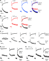
Figure 1.. NSF is involved in slow endocytosis at calyces
(A) Sampled calcium current (ICa) induced by depol20ms in a calyx of Held. (B) Sampled (single traces, left three traces) or averaged (right) capacitance changes induced by depol20ms (grey arrow head) at 4 - 10 min after break-in with a pipette containing a control solution (black, n = 11), 4 mM ATPγS (replacing ATP, red, n = 6) or 1 mM NEM (blue, n = 12). The Cm decay in Ctrl was fit mono-exponentially (τ: 10.6 s, gray). The amplitude of the ΔCmpeak for averaged traces was normalized (Avg/norm), and data were expressed as mean + SE every 5 – 10 s (applies to all other panels). Scale bars apply to panels B-D. (C) Similar to panel B, but with NSFmp (1 mM, black, n = 10) or NSFp (1 mM, red, n = 9). Traces on the left and middle are single traces, whereas traces on the right are normalized, averaged traces. (D) Similar to panel C, but with SNAPsp (1 mM, black, n = 7) or SNAPp (1 mM, red, n = 8). (E) The Ratedecay_n, ΔCm35s, ΔCmpeak, and QICa induced by depol20ms at 4 - 10 min after break in with a pipette containing the control solution (Ctrl, n = 11), ATPγS (4 mM, n = 6), NEM (1 mM, n = 12), NSFmp (1 mM, n = 10), NSFp (1 mM, n = 9), SNAPsp (1 mM, n = 7) or SNAPp (1 mM, n = 8). (F-J) Similar arrangement as panel A-E (including the calyx number), respectively, except that the stimulus was depol20msX10 and the ΔCm35s was replaced with ΔCm50s (E). The Cm decay in the Ctrl trace in B was fit bi-exponentially with τ of 1.4 s (weight: 29%) and 18.3 s, respectively (gray).
Figure 2.
Figure 2.. NSF knockout inhibits endocytosis at hippocampal synapses
(A-B) Sampled western blot of NSF, clathrin heavy chain (CHC), adaptor protein 2 α subunit (AP2), dynamin 1 (Dyn 1), and β-actin at day 0, day 4, and day 8 after Cre-4-OHT treatment to the NSFLoxP/LoxP hippocampal culture. (B) The NSF intensity (mean + s.e.m. from 8 cultures) recorded from the western blot at day 0, day 4, and day 8 after Cre-4-OHT treatment to the NSFLoxP/LoxP hippocampal culture. (C-D) The traces (C), Ratedecay_n (D) and ΔF/F (D) of SypH fluorescence (FSypH) changes (mean + s.e.m.) induced by Train20Hz in control (n = 20 experiments, wildtype, black), NSF−/− hippocampal boutons (n = 19 experiments, red), and NSF−/− boutons overexpressed with wildtype NSF (NSF−/−+NSF; blue; containing EBFP2 for recognition, n = 23) at 22–24 oC. Traces are also normalized and overlapped to show the block of the FSypH decay (C, right). *, p < 0.05; **, p < 0.01 (t test). (E-G) The traces (E, F), Ratedecay_n (G) and ΔF/F (G) of FSypH changes (mean + s.e.m.) induced by a 10 s AP train at 5 Hz (E, G) or 40 Hz (F, G) in control (5 Hz: 11 experiments; 40 Hz: 11 experiments; black) and in NSF−/− hippocampal boutons (5 Hz: 10 experiments; 40 Hz: 10 experiments; red) at 22–24 oC. Traces are also normalized and overlapped to show the block of the FSypH decay (E-F, right). Ratedecay_n and ΔF/F induced by AP20Hz are also included in panel G for comparison. (H-I) The traces (H), Ratedecay_n (I) and ΔF/F (K) of FSypH changes (mean + s.e.m.) induced by Train20Hz in control (n = 19 experiments, wildtype, black) and NSF−/− hippocampal boutons (n = 9 experiments, red) at 34–37 oC. Traces are also normalized and overlapped to show the block of the FSypH decay (H, right). (J-K) The traces (J), Ratedecay_n (K) and ΔF/F (K) of FSypH changes (mean + s.e.m.) induced by Train20Hz in wildtype cultures expressed with wildtype NSF (n = 14 experiments, +NSF, black) or NSFE329Q (n = 8 experiments, +NSFE329Q) at 22–24 oC. Traces are also normalized and overlapped to show the block of the FSypH decay (J, right).
Figure 3.
Figure 3.. NSF knock-out affects endocytosis examined with EM at hippocampal synapses.
(A) EM images of WT and NSF−/− hippocampal boutons at rest (Rest) and at 0 min (KCl), 3 min, and 10 min after 1.5 min 90 mm KCl application. For Rest, HRP was included for 1.5 min; for KCl application, HRP was included only during KCl application (see labels). (B-C) Number of HRP(+) vesicles (B) and the bulk endosome area (C) per square micrometer of synaptic cross-section are plotted versus the time before (Rest) and at 0 min (K+), 3 min, and 10 min after the end of KCl application in WT and NSF−/− hippocampal cultures (mean + SEM, each group was from 120–122 synaptic profiles from 18 mice (6 mice per experiment, 3 experiments). The temperature before fixation was 37°C. *: p<0.05; ***: p<0.001, t-test.
Figure 4.
Figure 4.. NSF is essential for mediating pre-Ω and fs-Ω pore closure in chromaffin cells
(A) Upper: setup drawing. The cell membrane, bath and vesicles are labelled with PHG (green), A655 (red) and FFN511 (yellow), respectively. ICa and Cm (capacitance) are recorded via a whole-cell pipette. Lower: Sampled ICa and Cm changes induced by depol1s. (B) PHG fluorescence (FPH), A655 fluorescence (F655) and sampled confocal images showing depol1s-induced (triangle) rapid (left), slow (middle) or large-size (right) pre-spot pore closure (pre-close) in chromaffin cells. FPH and F655 were normalized to the baseline. (C) FPH, F655, FFN511 fluorescence (FFFN), and confocal images showing close-fusion, stay- or shrink-fusion. (D) Western blot of NSF, clathrin heavy chain (CHC), dynamin 2 (Dyn 2), and β-actin in chromaffin cell cultures transfected with si-Ctrl or si-NSF. (E-G) The percentage of pre-spots undergoing depol1s-induced pre-close (E), the percentage of fusing vesicles undergoing close-fusion (F), and the 20–80% decay time of FFFN (G, indicating release time) in control (Ctrl, n = 25 cells), si-NSF transfection (n = 22 cells), si-NSF transfection plus wildtype NSF overexpression (si-NSF+NSF, 15 cells), control cells overexpressed with wildtype NSF (+NSF, 22 cells), or control cells overexpressed with NSFE392Q (+NSFE932Q, 24 cells). *: p < 0.05; **: p < 0.01(t test, compared to control).
Figure 5.
Figure 5.. NSF is essential for mediating slow, fast, ultrafast, and overshoot endocytosis in chromaffin cells
(A) Depol1s-induced ICa (mean + s.e.m., upper) and Cm (mean + s.e.m., lower) in chromaffin cells in control (Ctrl, 25 cells), si-NSF transfection (si-NSF, 22 cells), and si-NSF transfection plus wildtype NSF overexpression (si-NSF+NSF, 15 cells). Traces are also merged in the right. (B) Depol1s-induced ICa (mean + s.e.m., upper) and Cm (mean + s.e.m., lower) in chromaffin cells overexpressed with wildtype NSF (+NSF, 22 cells) or NSFE392Q (+NSFE392Q, 24 cells). Traces are also merged in the right. (C) Mean ICa (mean + s.e.m., upper) and Cm (mean + s.e.m., lower) induced by depol1s (gray triangle) in five groups of chromaffin cells (from left to right): Groupno-endo (decay < 30% ΔCm, 6 cells), Groupslow (endocytic τ > 6 s, 6 cells), Groupfast (τ: 0.6 – 6 s, 5 cells), Groupultrafast (τ < 0.6 s, 4 cells) and Groupovershoot (decay > 130% ΔCm, 4 cells) in control chromaffin cells. (D-E) Depol1s-induced ICa (mean + s.e.m., upper) and Cm (mean + s.e.m., lower) in chromaffin cells with ICa of 160 – 360 pA (left), 400 – 900 pA (middle), and 1000 – 1800 pA (right) in two conditions: (D) si-NSF transfection (left: 8 cells; middle: 7 cells; right: 7 cells); (E) NSFE392Q overexpression (left: 9 cells; middle: 8 cells; right: 7 cells).

Similar articles

References

    1. Littleton J.T., Chapman E.R., Kreber R., Garment M.B., Carlson S.D., and Ganetzky B. (1998). Temperature-sensitive paralytic mutations demonstrate that synaptic exocytosis requires SNARE complex assembly and disassembly. Neuron 21, 401–413. 10.1016/s0896-6273(00)80549-8. - DOI - PubMed
    1. Kuner T., Li Y., Gee K.R., Bonewald L.F., and Augustine G.J. (2008). Photolysis of a caged peptide reveals rapid action of N-ethylmaleimide sensitive factor before neurotransmitter release. Proc. Natl. Acad. Sci. U. S. A 105, 347–352. - PMC - PubMed
    1. Schweizer F.E., Dresbach T., Debello W.M., O’Connor V., Augustine G.J., and Betz H. (1998). Regulation of neurotransmitter release kinetics by NSF. Science 279, 1203–1206. - PubMed
    1. Kawasaki F., Mattiuz A.M., and Ordway R.W. (1998). Synaptic physiology and ultrastructure in comatose mutants define an in vivo role for NSF in neurotransmitter release. J Neurosci 18, 10241–10249. 10.1523/jneurosci.18-24-10241.1998. - DOI - PMC - PubMed
    1. He E., Wierda K., van Westen R., Broeke J.H., Toonen R.F., Cornelisse L.N., and Verhage M. (2017). Munc13–1 and Munc18–1 together prevent NSF-dependent de-priming of synaptic vesicles. Nat Commun 8, 15915. 10.1038/ncomms15915. - DOI - PMC - PubMed

Publication types

LinkOut - more resources