[Preprint]. 2025 Oct 15:2025.08.24.672028.
doi: 10.1101/2025.08.24.672028.

Reelin Controls the Directional Orientation, Apical Localization and Length of Primary Cilia in Principal Neurons in the Cerebral Cortex

Affiliations

Reelin Controls the Directional Orientation, Apical Localization and Length of Primary Cilia in Principal Neurons in the Cerebral Cortex

Sumaya Akter et al. bioRxiv. .

Abstract

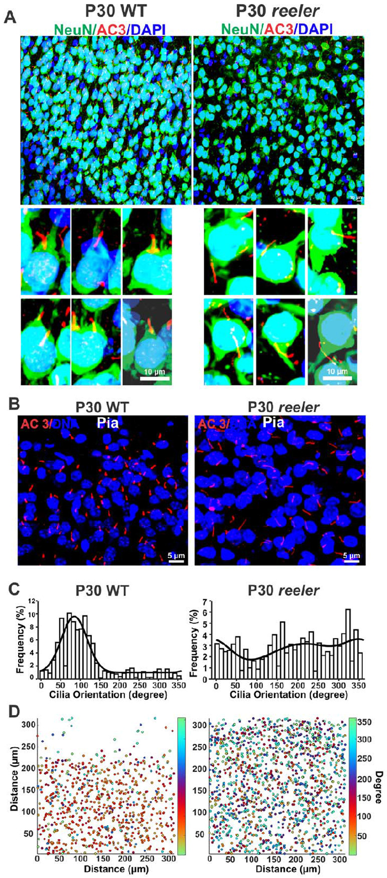
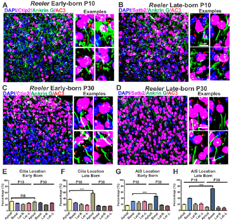
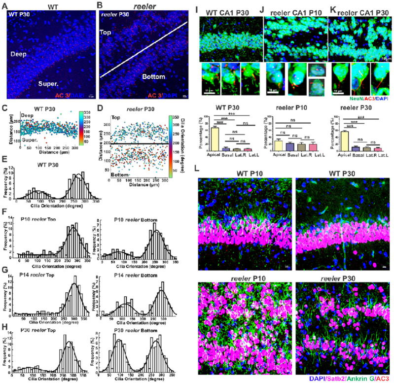
The primary cilia of pyramidal neurons in inside-out laminated regions orient predominantly toward the pia, reflecting reverse soma movement during postnatal neurodevelopment. However, the mechanisms underlying the directional cilia orientation are unknown. Here we show that the primary cilia of pyramidal neurons are localized near the base of the apical dendrites and aligned on the nuclear side opposite to the axon initial segment (AIS). However, this pattern is not observed in atypical pyramidal neurons in the deep neocortex, excitatory neurons in non-laminated regions, interneurons, or astrocytes, where cilia are irregularly positioned around the nuclei and lack preferred orientation. In Reelin-deficient mice (reeler), the directional orientation and apical location of cilia in late-born neocortical and CA1 neurons are disrupted. However, the initial impairments are partially corrected during postnatal development, along with a realignment of apical-basal orientation. In contrast, loss of Reelin drastically disrupts the directional orientation of cilia in early-born neocortical neurons and principal neurons in evolutionarily conserved cortical regions, which lack postnatal correction. Consistently, their cilia do not preferably localize to the apical side. Additionally, Reelin deficiency increases the cilia length of principal neurons across the cerebral cortex at a developmental stage when cilia stabilize in wild-type mice, but this effect is not observed in interneurons, astrocytes, or excitatory neurons in non-laminated regions. Together, Reelin controls the directional orientation, apical localization, and length of primary cilia in principal neurons in the cerebral cortex, underscoring the cilium as a key apical domain particularly prominent in late-born neurons.

Keywords: Apical-Basal Neuron Polarity; Cilia Orientation; Postnatal Neuron Positioning; Primary Cilia; Reelin; Reverse Movement.


Figures

Fig. 1.
Fig. 1.. Primary cilia of neocortical principal neurons are predominantly localized near the base of apical dendrites and aligned opposite to the AIS.
(A-C) Representative images showing cilia orientation and cilia location in the superficial layer. (A-B) Neurons marked by NeuN and Glutaminase antibodies. (C) (Left) A diagram showing how four quadrants (Apical, Basal, Lateral-Right and Lateral-Left) was defined in reference to the center of nucleus and apical dendrite. The location of cilia base in a quadrant was used to determine cilia location. (Right) Cilia predominantly localize to the apical quadrant in superficial neocortical neurons. (D-F) Late-born neurons cilia orientation and subcellular location. (D) Representative images showing cilia aligned opposite to AIS relative to the nuclei. (E) Cilia orientation histogram. (F) Cilia of late-born neurons were predominantly distributed to the apical quadrant. (G-I) Early-born neurons cilia orientation and location. (H) Cilia orientation histogram. (I) Cilia of early-born neurons were mostly distributed to the apical quadrant, while many still dispersed to other quadrants. E and H were fitted with one or two periodic normal distribution density functions, combined with a uniform distribution. One-way ANOVA tests were used to compare groups in cilia location.
Fig. 2.
Fig. 2.. Disrupted directional cilia orientation in the outer layer of the reeler neocortex.
(A-B) Primary cilia in the outer layer of the WT and reeler neocortices. WT cilia were mostly localized to the apical dendrites and oriented to the pia, while reeler cilia were irregularly distributed and lacked preferred orientation. (C) Histograms of cilia orientations in the outer layers of the reeler neocortex. WT cilia oriented toward the pia (peak at ~90°), while reeler cilia did not. Data were fitted with one or two periodic normal distribution density functions, combined with a uniform distribution. (D) Orientation map in the outer layers visualizing cilia orientation across the sampled fields, color-coded by angle (degrees). Data were pooled from multiple sections out of multiple reeler and WT brains.
Fig. 3.
Fig. 3.. Reeler late-born neurons exhibit a stronger postnatal rectification in apical-basal orientation than early-born neurons.
Reeler early-born neurons (A) and late-born neurons (B) at P10; early-born neurons (C) and late-born neurons (D) at P30. Primary cilia, AIS, early- and late-born neurons were labeled by AC3, Ankyrin G, CTIP2, and Satb2 antibodies, respectively. (E-H) cilia and AIS distribution in four quadrants of P10 and P30. At P10, mis-localized cilia and axon present in both early- and late born neurons. At P30, most late-born neurons’ cilia were localized to the apical quadrant and axons to the basal quadrant, while early-born neuron’s cilia were randomly distributed to four quadrants.
Fig. 4.
Fig. 4.. reeler late-born neocortical neurons are subject to corrections in cilia orientation and cilia apical location during postnatal development.
(A-D) reeler late-born neurons at P5 (A), P10 (B), P14 (C) and P30 (D). (i) Images of late-born neurons and primary cilia. (ii) Images of late-born neurons and cilia marked by NeuN and AC3 antibodies, respectively. NeuN staining display the apical dendrites. (iii) Representative images of individual neurons with apical dendrites and cilia. (iv) Cilia orientation of late-born neurons at different ages. Histograms of cilia orientation with fitting curves. (v) Cilia intracellular location in four quadrants at different ages. One-way ANOVA tests were used to compare groups.
Fig. 5.
Fig. 5.. reeler early-born neocortical neurons do not exhibit postnatal corrections in cilia orientation and apical location.
(A-B) cilia of reeler early-born neurons at P5 (A) and P30 (B). Cilia lack preferred orientation and apical location. Early-born reeler neurons at P5 (C-E) and P30 (F-H) were marked by a CTIP2 antibody. (C - F) Representative early-born neuron and cilia images. (D and H) Histogram of cilia orientation of early-born neurons. (E - H) Cilia intracellular location in four quadrants. One-way ANOVA tests were performed to compare groups. (I) Schematic diagrams depicting cilia orientation and intracellular location in neocortical principal neurons of WT and reeler at P30.
Fig. 6.
Fig. 6.. Reelin deficiency partially alters cilia orientation and cilia apical localization in CA1 pyramidal neurons.
(A-B) Representative images in WT (A) and reeler (B) CA1 regions at P30. reeler CA1 split “top” and “bottom”. (C-D) Cilia orientation map in WT (C) and reeler CA1 (D), visualizing cilia orientation across the sampled fields, color-coded by angle (degrees). (E) Histogram of cilia orientation in WT CA1 at P30. (F - H) Histograms showing cilia orientation in the top and bottom of reeler at P10 (F), P14(G), and P30 (H), respectively. (I-K) WT at P30 (I), reeler P10 (J) and reeler P30 (K). Cilia location in reeler CA1 neurons rectified during postnatal development from P10 to P30. Top: Representative CA1 images; Middle: individual neurons. Bottom: cilia location in four quadrants. ANOVA tests (L) WT CA1 neurons clearly exhibit a cilia-at-apical and axon-at-basal pattern at both P10 and P30. reeler CA1 neurons manifest altered localization pattern at P10 but had corrections at P30.
Fig. 7.
Fig. 7.. Disrupted cilia orientation and apical localization in the reeler piriform cortex.
WT (A) and reeler (B) piriform cortex of P30. Representative images of individual neurons are shown on the right. Histogram of cilia orientation in Layer II and III of WTs (C-D) and in outer and inner layers of reeler of P30 (E-F). (G) Percentage of cilia subcellular location in 4 quadrants. Cilia of WT neurons, but not reelers, primarily distribute to the apical quadrant.
Fig. 8.
Fig. 8.. Disrupted layering and ciliation pattern in the reeler DG and CA3.
Representative neuron and cilia images, marked by NeuN and AC3 antibodies, respectively. Data were collected from P30 brain samples. Histograms of cilia orientation and cilia intracellular location in WT and reeler are shown on the right. The pattern of cilia orientation and cilia intracellular location in WT DG and CA3 were not as strong or easily distinguishable as neocortical neurons. However, the loss of Reelin clearly altered the preferred apical location of cilia in DG and CA3.
Fig. 9.
Fig. 9.. Reelin deficiency promotes cilia elongation in principal neurons after P14 throughout the cerebral cortex.
(Left) Representative images of neuronal primary cilia (marked by an AC3 antibody) in different brain regions of WT and Reeler at P14 and P30. (Right) Bar graph showing cilia length of P14 and P30 WT and reeler mice. (A) CA1; (B) CA3; (C) Deep Neocortex; (D) Superficial Neocortex; and (E) Piriform Cortex. Two-tailed Student’s T-tests were performed to compare two groups.

References

    1. Alexander A., Herz J., and Calvier L.. 2023. Reelin through the years: From brain development to inflammation. Cell Rep. 42:112669. - PMC - PubMed
    1. Bangs F., and Anderson K.V.. 2017. Primary Cilia and Mammalian Hedgehog Signaling. Cold Spring Harb Perspect Biol. 9.
    1. Barbas H. 2015. General cortical and special prefrontal connections: principles from structure to function. Annu Rev Neurosci. 38:269–289. - PubMed
    1. Bishop G.A., Berbari N.F., Lewis J., and Mykytyn K.. 2007. Type III adenylyl cyclase localizes to primary cilia throughout the adult mouse brain. J Comp Neurol. 505:562–571. - PubMed
    1. Blackstad J.S., Osen K.K., and Leergaard T.B.. 2022. The fibro- and cyto-architecture demarcating the border between the dentate gyrus and CA3 in sheep (Ovis aries) and domestic pig (Sus scrofa domesticus). Hippocampus. 32:639–659. - PMC - PubMed

Publication types

LinkOut - more resources