[Preprint]. 2025 Aug 23:2025.08.20.671199.
doi: 10.1101/2025.08.20.671199.

Toggling of NKG2A expression drives functional specialization of iPSC-derived CAR NK cells

Affiliations

Toggling of NKG2A expression drives functional specialization of iPSC-derived CAR NK cells

Minoru Kanaya et al. bioRxiv. .

Abstract

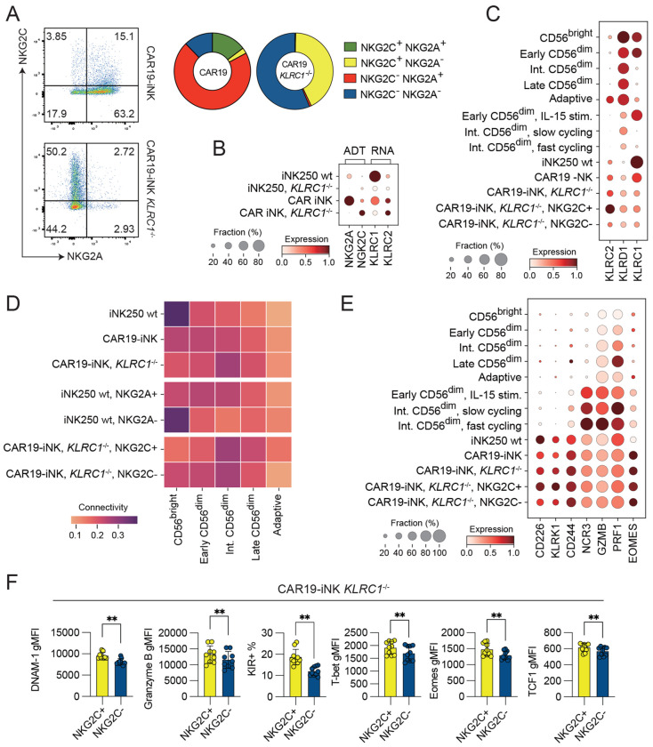
Induced pluripotent stem cell (iPSC)-derived natural killer (iNK) cells offer a promising platform for off-the-shelf immunotherapy against hematological malignancies. NK cell function is dynamically regulated through education driven by inhibitory receptors, including CD94/NKG2A and killer cell immunoglobulin-like receptors (KIR). However, the acquisition of inhibitory receptors in iNK cells and their role during differentiation and education remains poorly defined. In this study, we monitored receptor repertoires, transcriptional states, and functional responses in a range of genetically engineered iNK cell lines. Transcriptional reference mapping placed iNK cells close to cytokine-activated NKG2A+ CD56dim peripheral blood (PB) NK cells. Despite their early differentiation stage, iNK cells displayed a well-developed cytotoxic effector program, which was also reflected in high protein expression of Eomes, granzyme B, and activating receptors DNAM-1 and NKG2D. Acquisition of NKG2A by iNK cells was associated with a more differentiated transcriptional state and superior functional responses against a broad range of targets, including those expressing low to moderate levels of HLA-E, suggesting attenuated inhibitory signaling through NKG2A in iNKs. CRISPR knockout of β2-microglobulin (B2M) in iNK cells revealed that the functional potency of NKG2A+ iNK cells was independent of educating interactions with HLA-E in cis or trans. Finally, CRISPR-mediated ablation of NKG2A led to a spontaneous compensatory surface expression of CD94/NKG2C heterodimers, associated with enhanced IFN-γ production and cytotoxic activity against target cells with forced high expression of single-chain β2m-HLA-E-peptide trimers. Our results indicate an education-independent functional maturation of iNK cells, characterized by potent effector programs coupled with a favorable early-stage transcriptional profile.


Figures

Figure 1.
Figure 1.. Transcriptional reference mapping of iPSC-derived NK cells.
(A) Schematic of iNK cell generation and genetic engineering strategies, including CD19 CAR/IL-15/IL-15R transduction and KLRC1 knockout. (B) Schematic overview of peripheral blood NK (PB-NK) and IL-15 stimulated NK cell single-cell RNA sequencing datasets. (C-D) Mapping of iNK cells onto a PB-NK reference. (E) Partition-based graph abstraction (PAGA) depicting connectivity between engineered iNK subsets and PB-NK cells. (F-G) Transcriptional states, regulatory programs and comparison of differentiation sages of iNK subsets compared to PB-NK subsets. n = 2-12.
Figure 2.
Figure 2.. Functional superiority of NKG2A+ iNK cells.
(A) Flow cytometric analysis of NKG2A and NKG2C expression in resting and expanded PB-NK cells and iNK lines (iNK121 wt, iNK250 wt, and CAR19-iNK). (B-E) Degranulation (CD107a) and IFN-γ production assays demonstrate enhanced cytotoxicity of NKG2A+ subsets compared to NKG2A− subsets in PB-NK and iNK lines against K562 (B-D) and Nalm-6 targets (E). (F) Cytotoxicity of NKG2A+ compared to NKG2A− iNK cells against K562 target cells. (G) Phenotypic characterization of resting PB-NK, expanded PB-NK and iNK lines (iNK205, CAR19-iNK) stratified by NKG2A expression. (H-J) Granzyme B, perforin, DNAM-1, and Eomes expression levels in NKG2A+ CAR19-iNK cells.. Data are represented as mean (SD). Significance was calculated using a Wilcoxon matched-pairs signed rank test (B-C, H-I) or a Friedman test followed by Dunn’s multiple comparison test (D-E). p values: * < 0.05, ** < 0.01, *** < 0.001, **** < 0.0001. n = 2-13.
Figure 3.
Figure 3.. Education-independent functional maturation of NKG2A+ iNK cells.
(A-B) Schematic overview and surface expression of HLA-ABC and HLA-E in wild-type (iNK wt) and β2-microglobulin-deficient (iNK B2M−/−) iNK cells. (C) Distribution of NKG2A and NKG2C subsets in the iNK121 B2M−/− and iNK250 B2M−/− cell lines. (D-E) Functional assays (CD107a, IFNγ) of iNK121 B2M−/− and iNK250 B2M−/− cells against K562 target cells, stratified by NKG2A expression. Data are represented as mean (SD). Significance was calculated using a Wilcoxon matched-pairs signed rank test. p values: * < 0.05, ** < 0.01, *** < 0.001, **** < 0.0001. n = 4-12.
Figure 4.
Figure 4.. HLA-E levels modulate iNK cell functionality.
(A-B) Flow cytometric analysis of HLA-ABC and HLA-E expression in target cells, including K562 and Nalm-6 cells with varying HLA-E expression levels. (C) Degranulation (CD107a) and IFNγ assays demonstrate of resting PB-NK, iNK250 and iNK-CAR19 cells to targets with supraphysiological HLA-E expression. (D) Relative response rate calculations of NKG2A+ and NKG2A− subsets against targets with varying levels of HLA-E expression (E) Degranulation (CD107a) and IFNγ production of CAR19-iNK against Nalm-6 cell lines expressing varying HLA-E levels. (F-G) Functional responses of CAR19-iNK cells expressing or lacking NKG2A against Nalm-6 HLA-E variants. Data are represented as mean (SD). Significance was calculated using a Friedman test followed by Dunn’s multiple comparison test. p values: * < 0.05, ** < 0.01, *** < 0.001, **** < 0.0001. n = 7-12.
Figure 5.
Figure 5.. Adaptive reprogramming of CAR19-iNK cells following KLRC1 knockout.
(A-C) NKG2C expression at the protein (A-B) and transcript level (B-C) after CRISPR knockout of KLRC1 in iNK-CAR19 cells. (C-D) Mapping of iNK cell lines onto a PB-NK reference map. (E-F) Expression of activating receptors, effector molecules, and transcription factors in reference populations and CAR19-iNK KLRC1−/− cells stratified by NKG2C expression. Data are represented as mean (SD). Significance was calculated using a Wilcoxon matched-pairs signed rank test. p values: * < 0.05, ** < 0.01, *** < 0.001, **** < 0.0001. n = 2-12.
Figure 6.
Figure 6.. Enhanced cytotoxicity of CAR19-iNK KLRC1−/− cells against HLA-E-high targets.
(A-B) Degranulation (CD107a) and IFNγ production assays comparing CAR19-iNK KLRC1−/− cells to wild-type CAR19-iNK cells against K562 (A) and Nalm-6 cells (B) with varying HLA-E levels. (C) Relative response analyses of KLRC1 knockout cells against target cells with varying HLA-E expression. (D-E) Long-term killing assays of CAR19-iNK variants cells against Nalm-6 target cells expressing varying levels of HLA-E. Data are represented as mean (SD). Significance was calculated using a Wilcoxon matched-pairs signed rank test (A-B) or a Friedman test followed by Dunn’s multiple comparison test (C). p values: * < 0.05, ** < 0.01, *** < 0.001, **** < 0.0001. n = 5-12.

References

    1. Saetersmoen M.L., Hammer Q., Valamehr B., Kaufman D.S. & Malmberg K.J. Off-the-shelf cell therapy with induced pluripotent stem cell-derived natural killer cells. Semin Immunopathol 41, 59–68 (2019). - PubMed
    1. Themeli M., Riviere I. & Sadelain M. New cell sources for T cell engineering and adoptive immunotherapy. Cell Stem Cell 16, 357–366 (2015). - PMC - PubMed
    1. Zhu H. et al. Pluripotent stem cell-derived NK cells with high-affinity noncleavable CD16a mediate improved antitumor activity. Blood 135, 399–410 (2020). - PMC - PubMed
    1. Zhu H. et al. Metabolic Reprograming via Deletion of CISH in Human iPSC-Derived NK Cells Promotes In Vivo Persistence and Enhances Anti-tumor Activity. Cell Stem Cell 27, 224–237 e226 (2020). - PMC - PubMed
    1. Cichocki F. et al. iPSC-derived NK cells maintain high cytotoxicity and enhance in vivo tumor control in concert with T cells and anti-PD-1 therapy. Science translational medicine 12 (2020). - PMC - PubMed

Publication types