[Preprint]. 2025 Aug 22:2025.08.22.671264.
doi: 10.1101/2025.08.22.671264.

Predictive encoding of auditory sequences in the human prefrontal cortex

Affiliations

Predictive encoding of auditory sequences in the human prefrontal cortex

Olgerta Asko et al. bioRxiv. .

Abstract

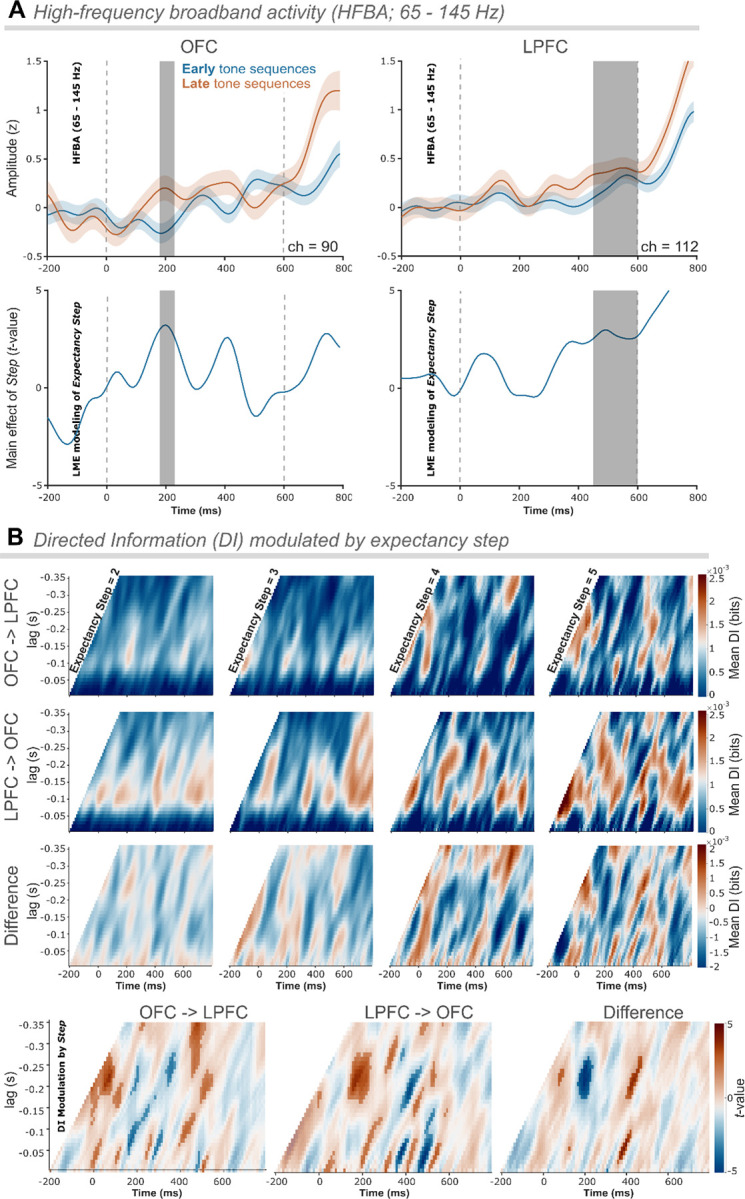
Humans extract regularities from the environment to form expectations that guide perception and optimize behavior. Although the prefrontal cortex (PFC) is central to this process, the relative contributions of orbitofrontal (OFC) and lateral PFC (LPFC) remain unclear. Here, we show that the brain tracks sound regularities in an auditory deviance detection task to predict when a target deviant will occur. Intracranial EEG in epilepsy patients reveals prefrontal engagement, with earlier expectancy-related modulation in OFC and later modulation in LPFC. Connectivity analyses indicate bidirectional and asymmetrical expectancy-related information exchange between the two areas, with a first lead by OFC, consistent with its role in initiating predictive encoding. Converging causal evidence shows that OFC lesions abolish sensitivity to expectancy, whereas LPFC lesions yield only modest effects not significantly different from controls. Together, these results provide electrophysiological and causal evidence for distinct, temporally organized contributions of prefrontal subregions to predictive processing.

Keywords: Contingent Negative Variation (CNV); EEG; High-Frequency Broadband Activity (HFBA); SEEG; anticipation; auditory perception; deviance detection; expectation; frontal lobe lesion; lateral prefrontal cortex; orbitofrontal cortex; prediction.


Conflict of interest statement

Competing interests The authors declare no competing interests.

Figures

Figure 1.
Figure 1.. Illustration of task design, lesion reconstruction in patients with frontal damage, and depth electrode coverage in patients with epilepsy.
(A) Auditory local-global deviance detection task. Sequences of 50 ms-duration tones were presented with a fixed stimulus onset asynchrony (SOA) of 150 ms and a uniformly jittered inter-trial interval (ISI) of 700–1000 ms. Each block began with a habituation phase, consisting of 20 identical tone sequences, which would occur commonly throughout the rest of the block. Then, in the remaining part of the block, deviant sequences (25%) were pseudorandomly interspersed among the standards. In regular blocks (XX), standard sequences (75%) consisted of five repetitions of the same tone. They were interspersed with deviant sequences where the fifth sound was either different in frequency type (12.5%) or was shorter (12.5%). In irregular blocks (XY), standard sequences (75%) had a fifth sound differing in frequency type, interspersed with deviant sequences with five repetitions of the same tone (12.5%) or shorter sequences of four identical tones (12.5%). In omission blocks (XO), standard sequences consisted of four repetitions of the same tone. They were mingled with deviant sequences with five repetitions of the same tone (12.5%) or with the fifth sound at a different frequency (12.5%). The expectancy step number of each sequence was defined with respect to the numbering since the last deviant sequence. Reaction times. Increased speed of global deviant’s detection with expectancy. Violin plots depicting mean reaction times (RT) in milliseconds (ms) across the four expectancy steps (2–5) in the RT version of the task conducted with healthy participants (BEH group). Each violin plot represents the distribution of RTs for each step, with individual participant data points connected by gray lines. As the steps increased, a trend towards faster RTs was observed, indicating enhanced response efficiency with increased expectancy. (B) Reconstructed lesions in patients with OFC and LPFC damage. Lesion overlap for orbitofrontal cortex (OFC: top; n = 12) and lateral prefrontal cortex (LPFC: bottom; n = 10) patients. Aggregate lesion overlay maps per group in axial view. The color code (from 0 to 100%) indicates the percentage of shared lesion coverage across patients. The redder the color, the greater the lesion overlap. Neurological convention was used with the right side of the brain being displayed on the right side of the image and vice versa. (C) Intracerebral depth electrodes in patients with drug-resistant epilepsy. Normalized space projection of the 202 recorded electrodes onto the 3D glass brain reconstruction of the orbital (red surface; electrodes = 90) and lateral (blue surface; electrodes = 112) PFC. The color-coding of electrodes indicates the individual patients. Note that, by chance, there were more patients with right- than left-hemisphere electrode coverage during the study period (right: n = 6; left: n = 1).
Figure 2.
Figure 2.. Scalp-recorded contingent negative variation (CNV) was modulated by global deviant expectancy in healthy adults and was significantly reduced in patients with lesions to the orbitofrontal cortex (OFC).
(A) Event-related potentials. On the top, healthy control participants’ (Controls) grand average CNV waveforms at two exemplary electrodes, AF8 (left) and Cz (right). CNV waveforms elicited by early tone sequences (expectancy step = 2) are in blue, and in brown for late tone sequences (expectancy step = 4 and 5). In the middle, orbitofrontal cortex (OFC) lesion patients’ grand average CNV waveforms are shown at the same electrodes and for the same task conditions. At the bottom, lateral prefrontal cortex (LPFC) lesion patients’ grand average CNV waveforms are depicted at the same electrodes for the same conditions. Dashed lines indicate the average linear regression from where CNV slopes were extracted. Vertical lines denote tone onsets. Scalp topographies of voltage averaged in the 600 ms time window from the onset of the first tone to the onset of the fifth tone for early and late tone sequences show a fronto-central negativity maximum, which is more negative for late compared to early tone sequences. (B) Mean CNV slope values. Violin plots of mean slope value over expectancy steps (2–5) obtained from fronto-central electrodes (i.e., AF7, AF8, Fz, FC3, FCz, FC4, C3, Cz, and C4) for the Controls and the OFC and LPFC lesion groups. Lines connect slope values (points) from the same participant. (C) Linear mixed-effects model. On the left, scalp topographies show the t-values distribution of the expectancy step effect on the CNV slope for the Controls, the OFC, and the LPFC patients, respectively, when modeled independently. To the right, the scalp topographies illustrate the effect of group (top) and the interaction effect of expectancy step and group (bottom) for OFC vs. Controls (left), LPFC vs. Controls (middle), and OFC vs. LPFC (right). Below the expectancy step effects, participants’ t-values calculated for fronto-central electrodes (see B) obtained with individual linear models.
Figure 3.
Figure 3.. Intracerebral high-frequency broadband activity (HFBA) recorded from orbitofrontal and lateral PFC is modulated by deviance expectancy.
(A) High-frequency broadband activity (HFBA; 65 – 145 Hz). Average HFBA signals recorded from orbitofrontal (OFC; left) channels (n = 90) and lateral prefrontal (LPFC; right) channels (n = 112). HFBA elicited by early tone sequences (expectancy step = 2) are in blue, and HFBA signals elicited by late tone sequences (expectancy step = 4 and 5) are in brown. Gray area denotes a significant modulation of expectancy step in the linear mixed-effects (LME) modeling. Bottom: Temporal LME modeling t-values time course (−200 – 800 ms) with marked significant segments (gray vertical bar) of expectancy step modulation for OFC (left) and LPFC (right). (B) Directed Information (ID) modulated by expectancy step. The color scale reflects the strength of information flow, with warmer colors indicating higher DI values. First row: average DI flow between OFC and LPFC channel pairs (n = 1482); Second row: average DI flow between LPFC and OFC channel pairs (n = 1482); Third row: average difference (OFC → LPFC minus LPFC → OFC) DI flow. Bottom row: LME modeling t-values time course (− 200 – 800 ms) showing the modulation of DI flow by expectancy step between OFC and LPFC (left), between LPFC and OFC (middle), and the DI difference (OFC → LPFC minus LPFC → OFC; right). Positive DI differences indicate OFC lead (brown), whereas negative values indicate LPFC lead (blue). Significant modulation of the information flow by expectancy step (statistically significant t-values, uncorrected for left and middle plots, FDR-corrected for right plot) are represented by the opaque colored areas, while non-significant results are shown as transparent areas.

Similar articles

References

    1. Rao RP, Ballard DH. Predictive coding in the visual cortex: a functional interpretation of some extra-classical receptive-field effects. Nature neuroscience. 1999;2(1):79–87. - PubMed
    1. Clark A. Whatever next? Predictive brains, situated agents, and the future of cognitive science. Behavioral and brain sciences. 2013;36(3):181–204. - PubMed
    1. Friston K. A theory of cortical responses. Philosophical transactions of the Royal Society B: Biological sciences. 2005;360(1456):815–836. - PMC - PubMed
    1. Friston K. The free-energy principle: a unified brain theory? Nature reviews neuroscience. 2010;11(2):127–138. - PubMed
    1. Daw ND, Gershman SJ, Seymour B, Dayan P, Dolan RJ. Model-based influences on humans’ choices and striatal prediction errors. Neuron. 2011;69(6):1204–1215. - PMC - PubMed

Publication types

LinkOut - more resources