Review
. 2025 Aug 14:13:1650885.
doi: 10.3389/fcell.2025.1650885. eCollection 2025.

Stem cell therapy offers new hope for the treatment of Alzheimer's disease

Affiliations
Review

Stem cell therapy offers new hope for the treatment of Alzheimer's disease

Guodong He et al. Front Cell Dev Biol. .

Abstract

Alzheimer's disease (AD) is a progressive neurodegenerative disorder primarily characterized by memory impairment and cognitive decline, for which no curative treatment is currently available. Existing therapeutic strategies, such as cholinesterase inhibitors and N-methyl-D-aspartate (NMDA) receptor antagonists, can only provide limited symptomatic relief and fail to halt disease progression. In recent years, stem cell therapy has emerged as a promising approach for AD due to its multifaceted mechanisms of action. The therapeutic effects of stem cells in AD are mainly attributed to their ability to differentiate into functional neurons or glial cells, thereby replacing damaged cells and repairing neural networks. In addition, stem cells secrete neurotrophic and anti-inflammatory factors that contribute to the improvement of the brain microenvironment. Furthermore, they can regulate neuroinflammation, promote the clearance of β-amyloid (Aβ) deposits, and suppress neuroinflammation, thus potentially slowing disease progression. However, several challenges remain, including low cell survival rates, immune rejection, tumorigenic risks, and difficulties in crossing the blood-brain barrier. Looking ahead, the integration of advanced technologies such as organoid models, gene editing, artificial intelligence, and multi-omics approaches may drive substantial progress in the clinical translation of stem cell therapies for AD. Although still in its early stages, the future of this therapeutic strategy holds great promise.

Keywords: alzheimer’s disease; amyloid-β clearance; paracrine and immunomodulatory mechanisms; regenerative therapy; stem cells.


Conflict of interest statement

The authors declare that the research was conducted in the absence of any commercial or financial relationships that could be construed as a potential conflict of interest.

Figures

FIGURE 1
FIGURE 1
The pathogenesis of Alzheimer’s disease.
FIGURE 2
FIGURE 2
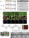
Selection of the optimal media for triculture of human iPSC-derived neurons, astrocytes, and microglia (A) Schematic of the iPSC differentiation protocols for neurons (iNs), astrocytes (iAs), and microglia (iMGs). iNs and iAs were generated by lentiviral expression of lineage-specific transcription factors, while iMGs were derived via a hematopoietic precursor (HPC) stage using non-viral methods. Cryopreservation days are indicated; each cell type was fully differentiated before switching to triculture media (TCM). Key abbreviations: KSR, knockout serum replacement; N2B, neu robasal with N2/B27; EM, expansion medium; FGF, fibroblast growth factor medium. (B) Table summarizing the composition of each cell-type-specific media and TCM. (C–E) Representative immunostaining of neurons (C), astrocytes (D), and microglia (E) maintained in either their respective media or TCM. Markers shown include TUJ1/NeuN for neurons, GFAP/S100B for astrocytes, and IBA1/INPP5D for microglia. Scale bars, 50 μm. (F) Timeline of the triculture workflow. Cryopreserved iNs, iAs, and iMGs were thawed and matured separately; astrocytes and microglia were sequentially plated onto neuron cultures on days 20 and 21 and then co-cultured for 3–6 days. The days in bold refer to the start of thawing the first stock of cryopreserved cells, and the non-bolded days refer to the day of differentiation for each cell type. (G) Bar plot showing the relative percentages of NeuN + (neurons), IBA1 + (microglia), and CD44 + (astrocytes) cells at day 27, determined from immunostaining (n = 2 genetic backgrounds, three differentiations, and two wells per differentiation). Error bars represent standard error. A representative field of view (FOV) is shown, with six FOVs per well analyzed by blinded quantification. Scale bar, 200 μm. (H) Representative triculture images (3–6 days of co-culture) labeled for CD44 (astrocytes), IBA1 (microglia), and TUJ1 (neurons). Scale bars, 100 μm. (I) Western blot of tricultures at days 3 and 6, probed for INPP5D, IBA1, TAU5, CD44, and GAPDH, confirming the presence of all 3 cell types (Lish et al., 2025)
FIGURE 3
FIGURE 3
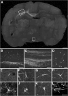
Incorporation of ES cell-derived neurons into the developing rat brain. (A–K) Engrafted donor cells identified by their EGFP fluorescence (E,G–K) or immunofluorescence with an antibody to EGFP (A–D,F) generate a variety of neuronal phenotypes. A, Twenty days after transplantation into the ventricle of E16.5 rats, the cells formed intraventricular clusters and migrated as single cells into various host brain regions. (B–D) Higher power microphotographs of areas indicated in (A) depicting incorporation into neocortex(B)and hypothalamus(D). Donor-derived cortical neurons were found to extend long axons into the corpus callosum (C). (E–K) Confocal microscopy and digital reconstruction revealed that the transplanted cells adopt a variety of morphologies, including simple bipolar cells resembling young migratory neurons (E, neocortex), complex phenotypes mimicking principal pyramidal neurons of the hippocampus (F, CA1pyramidalcelllayer), and multipolar cell types (G,H) neocortex; (I) septum; (J), thalamus; (K), tectum). (L) Immunofluorescence analysis with an antibody to nestin depicts engrafted cells with immature, elongated phenotypes characteristic of migratory precursor cells. The arrow points to the mouse-specific DNA in situ hybridization signal used for donor cell identification (tectum, confocal analysis). Scale bars: (A) 1 mm; (B) 200 m; (C) (D) 100 m; (E–L) 50 m (Wernig et al., 2004b).
FIGURE 4
FIGURE 4
Oxidative stress in hippocampal neurons exposed to AβOs in the absence or presence of MSCs. Photomicrographs showing DCF fluorescence (green) in hippocampal neurons exposed to vehicle (A–D) AβOs (500 nM) for (E–H) or H2O2 (100 M) for 10min (I–L) in the absence or presence of MSCs, as indicated. Scale bar, 100 m. Images were acquired on a Nikon Eclipse TE300 epifluorescence microscope with a 20 objective. Corresponding bright-field images are shown beside each fluorescence image. (M–O) quantification of integrated DCF fluorescence intensity normalized by the total number of cells. Panels show integrated fluorescence for AβO-exposed neurons (M) H2O2-exposed neurons (N) or MSCs cocultured with hippocampal neurons and exposed to vehicle or AβOs, compared with hippocampal neurons alone (O). Data are represented as mean S.E. (error bars) (n 6independentcultures, with triplicate coverslips in each experimental condition); *, p 0.05; two-way ANOVA followed by Tukey’s post hoc test; RU, relative units (de Godoy et al., 2018)

Similar articles

References

    1. Acharya P., Choi N. Y., Shrestha S., Jeong S., Lee M. Y., Brain O. (2024). Brain organoids: a revolutionary tool for modeling neurological disorders and development of therapeutics. Biotechnol. Bioeng. 121 (2), 489–506. 10.1002/bit.28606 - DOI - PMC - PubMed
    1. Ahmed T., Zahid S., Mahboob A., Farhat S. M. (2017). Cholinergic system and post-translational modifications: an insight on the role in Alzheimer's disease. Curr. Neuropharmacol. 15 (4), 480–494. 10.2174/1570159x14666160325121145 - DOI - PMC - PubMed
    1. Al Abbar A., Ngai S. C., Nograles N., Alhaji S. Y., Abdullah S. (2020). Induced pluripotent stem cells: reprogramming platforms and applications in cell replacement therapy. BioResearch open access 9 (1), 121–136. 10.1089/biores.2019.0046 - DOI - PMC - PubMed
    1. Allen S. J., Watson J. J., Shoemark D. K., Barua N. U., Patel N. K. (2013). Gdnf, ngf and bdnf as therapeutic options for neurodegeneration. Pharmacol. and Ther. 138 (2), 155–175. 10.1016/j.pharmthera.2013.01.004 - DOI - PubMed
    1. Almkvist O., Graff C. (2021). The apoe Ε4 allele affects cognitive functions differently in carriers of app mutations compared to carriers of Psen1 mutations in autosomal-dominant Alzheimer's disease. Genes (Basel) 12 (12), 1954. 10.3390/genes12121954 - DOI - PMC - PubMed

LinkOut - more resources