. 2025 Aug 14:15:1643576.
doi: 10.3389/fcimb.2025.1643576. eCollection 2025.

Expanding structural insights into DNA packaging apparatus and endolysin LysSA05 function of Epsilon15 bacteriophage

Affiliations

Expanding structural insights into DNA packaging apparatus and endolysin LysSA05 function of Epsilon15 bacteriophage

Muhammad Saleem Iqbal Khan et al. Front Cell Infect Microbiol. .

Abstract

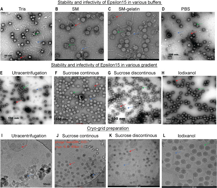
The rising prevalence of multidrug-resistant (MDR) foodborne pathogens, particularly Salmonella spp., necessitates alternative antimicrobial solutions. Phage therapy offers a promising solution against MDR Gram-negative infections; however, its clinical application is constrained by the presence of endotoxins, residual cellular debris, the risk of horizontal gene transfer by temperate phages, and an incomplete understanding of how phage structural integrity influences infectivity and enzyme function. In this study, we present a structural and functional analysis of temperate bacteriophage Epsilon15 (ϵ15), focusing on its DNA packaging and injection machinery, along with characterization of the dual-acting endolysin LysSA05. Iodixanol-purified virions suspended in phosphate-buffered saline (PBS), under conditions optimized to preserve virion stability, were analyzed using graphene oxide (GO)-supported cryo-electron microscopy. This approach resolved the full asymmetric architecture of ϵ15, revealing a detailed internal nucleic acid organization with at least eight concentric layers radially and approximately 28 axially compacted layers within the capsid. The DNA packaging machinery, comprising the core, portal, and hub, was resolved at high resolution, including a 42 nm-long and 18 nm-wide injection channel anchored by a dodecameric portal complex visualized at ~7 Å resolution. Concurrently, we characterized LysSA05, a dual-acting endolysin harboring a glycoside hydrolase 19 (GH19) catalytic domain accommodating peptidoglycan (PG) residues N-acetylmuramic acid (NAM) and N-acetylglucosamine (NAG) through structural docking, indicating plausible binding interactions that promote hydrolysis support vector machine (SVM), random forest (RF), discriminant analysis (DA), artificial neural network (ANN) and physicochemical scanning identified an amphipathic helix (residues 59-112) with predicted antimicrobial peptide (AMP)-like properties. Biochemical validation confirmed that LysSA05 destabilizes lipopolysaccharides (LPS) and permeabilizes the outer membrane of Gram-negative bacteria independently of permeabilizers, with enhanced efficacy observed upon co-treatment with Ethylenediaminetetraacetic acid (EDTA) or citric acid. In summary, our findings elucidate key structural features of ϵ15 relevant to infection and genome delivery, while positioning LysSA05 as a promising enzybiotic candidate against MDR Gram-negative pathogens.

Keywords: antimicrobial peptides; bacteriophage; cryo-electron microscopy; electron microscopy; endolysin; multidrug-resistant bacteria.


Conflict of interest statement

The authors declare that the research was conducted in the absence of any commercial or financial relationships that could be construed as a potential conflict of interest.

Figures

Figure 1
Figure 1
Assessment of ϵ15 virion stability under different purification conditions via EM/Cryo-EM grid preparation. (A–C) Negative-staining EM of ϵ15 particles purified using different buffer systems: (A) Tris, (B) SM buffer, (C) SM-gelatine, and (D) PBS. (E–H) Negative-staining EM of ϵ15 particles following density gradient centrifugation using: (E) ultracentrifugation, (F) continuous sucrose, (G) discontinuous sucrose, (H) discontinuous iodixanol gradients (I–L) Cryo-EM of ϵ15 virions vitrified on GO-coated grids following purification by: (I) ultracentrifugation, (J) continuous sucrose, (K) discontinuous sucrose, and (L) discontinuous iodixanol gradients. Red arrows indicate broken particles; green arrows denote intact virions; blue arrows highlight background debris. Scale bars are indicated 50, 100 or 200 nm in each EM panel.
Figure 2
Figure 2
Cryo-EM grid support evaluation and asymmetric reconstruction of the genome packaging machinery in ϵ15. (A) Cryo-EM micrographs of ϵ15 particles vitrified on a standard holey carbon grid, exhibiting strongly preferred orientation. (B) Cryo-EM micrographs of ϵ15 prepared on a perforated grid coated with a GO, demonstrating improved particle dispersion and orientation. (C) Icosahedral 3D reconstruction of the capsid, revealing the symmetric protein shell architecture. (D) Asymmetrical reconstruction of the full virion, capturing the tail complex anchored at a unique capsid vertex. (E) Cross-sectional view of the asymmetric map, with radial coloring highlighting concentric layers of packaged genomic DNA. (F) Cutaway view of the full reconstruction, illustrating the ordered DNA arrangement and the portal-tail complex. The schematic outlines three main components: core, portal, and hub regions (~42 nm total length, ~18 nm core width). (G) Side view of the portal-tail complex density map, shown in both intact and exploded views corresponding to the structural elements in panel (H) Focused 7 Å reconstruction of the portal region, revealing a dodecameric ring composed of 12 subunits critical for DNA translocation. Red arrow: the tail of ϵ15 aligned with the icosahedral axis.; Blue arrow: the tail of ϵ15 positioned at the side of the icosahedron. Scale bar: 200 nm for panels (A, B).
Figure 3
Figure 3
Structural features and molecular docking of LysSA05 endolysin with peptidoglycan ligands NAM and NAG. (A) Domain architecture of LysSA05 endolysin, highlighting the glycosidase family 19 (GH19) catalytic domain (pink) and the peptidoglycan-binding domain (green), along with predicted α-helical segments. (B) Ribbon diagram of the predicted 3D structure of LysSA05, showing domain organization and two prominent groove loops. (C) Superimposition of the predicted LysSA05 structure with a homologous template (PDB: 4OK7), shown in blue. (D) 2D chemical structure of NAM. (E) Surface representation of LysSA05 with NAM-binding cavity highlighting in yellow. (F) NAM docked into catalytic groove of LysSA05 (cartoon view); key interactions are shown as dashed lines. (G) Zoomed-in view of the NAM binding pocket, showing hydrogen bonds and hydrophobic contacts stabilizing the interaction. (H) 2D chemical structure of NAG. (I) Surface representation of LysSA05 with the NAG-binding pocket highlighted in blue. (J) NAG docked into LysSA05 cavity, visualizing ligand–protein interactions. (K) Close-up of the NAG binding site; showing hydrogen bonds are represented by green bashed lines, while red spokes denote hydrophobic interaction, with bond distance shown in angstrom Å.
Figure 4
Figure 4
AMP prediction from LysSA05 using machine learning and physicochemical profiling. (A) Preliminary-HeliQuest analysis showing net charge (z) and hydrophobic moment (µH) of predicted helical segments. (B–C) Helical wheel projections of top two amphipathic helices, illustrating residue polarity and spatial distribution. (D–G) Distribution of AMP and NAMP prediction across helix position based on four machine learning classifiers: SVM, RF, DA, and ANN. (H–J) AMP probabilities scores for predicted segments; AMP-classified helices consistently show significantly higher probability across all the models. (K) Heatmap representing AMP probabilities scores across the LysSA05 sequence, highlighting regions with high AMP potential. (L) Venn diagram illustrating the overlap of AMP predictions among SVM, RF, and DA classifiers. (M) Top- ranked AMP helices predicted by SVM, RF, and DA with probability scores ≥ 0.8. (N) Final selection of high-confidence AMP helices in LysSA05 identified across the three classifiers (SVM, RF, DA), each with AMP probability ≥ 0.8.
Figure 5
Figure 5
Amphipathic properties and α-helical structures of top AMP-like peptides derived from LysSA05 endolysin. (A–D) Helical wheel projections and physicochemical parameters for four top-ranking peptides (A) 59–78, (B) 67–86, (C) 68–87, and (D) 69–88 generated using HeliQuest. Each projection illustrates the distribution of residues around the helical axis, revealing distinct amphipathic separation: polar/charged residues (blue/purple) cluster on the solvent-exposed face, while hydrophobic residues (yellow/grey) align on the membrane-interacting face. Accompanying boxes summarize key parameters: hydrophobicity (H), hydrophobic moment (µH), and net charge (z). The 3D ribbon diagram (left panels) shows predicted α-helical confirmation of each peptide, highlighting their structural potential for membrane insertion and antimicrobial activity.
Figure 6
Figure 6
Expression optimization and purification of recombinant LysSA05. (A) Schematic diagram illustrating the cloning of gp05 into the pET-28a(+) vector using NcoI and XhoI restriction sites. (B–D) SDS-PAGE analysis of expression optimization in E. coli Rosetta under varying conditions: (B) 30°C with 1 mM IPTG; (C) 37°C with 0.5 mM IPTG; (D) 37°C with 1 mM IPTG. A distinct ~25 kDa band corresponding to His6-tagged LysSA05 is observed in lane 4 (Rosetta, 37°C, 1 mM IPTG), indicating optimal expression. (E) Purification of LysSA05 from E. coli BL21 showing low yield after Ni-NTA affinity chromatography across elution fractions 3-6, compared to control (lanes 1, 2). (F) Purification from Rosetta strain showing high expression and high >90% purity of the ~25 kDa protein across elution fractions 5-9. Lane 1: non-induced control; Lane 2: supernatant after centrifugation; Lane 3: soluble fraction post-sonication; Lane 4: total cell lysate post-sonication.
Figure 7
Figure 7
Characterization of LysSA05 LPS-degrading activity and enhancement of antibacterial activity by OMPs. (A) SDS-PAGE and silver staining demonstrating LPS degradation in E. coli C following treatment with LysSA05. (B) SDS-PAGE and silver staining showing LPS degradation in S. anatum treated with LysSA05. (C) CFU reduction assay indicating the direct antibacterial effect of LysSA05 against E. coli C and S. anatum, with log reductions of approximately 2.0 and 1.3, respectively, after 5 h of incubation. (D, E) Synergistic effect of various EDTA concentrations (mM) in combination with LysSA05 against E. coli C and S. anatum. (F, G) Synergistic effect of different citric acid concentrations (mM) in combination with LysSA05 against E. coli C and S. anatum.
Figure 8
Figure 8
Antimicrobial activity of LysSA05 against clinical isolates of E. coli and Salmonella. (A). Bactericidal effect of LysSA05 (100 μg/ml) against logarithmic-phase E. coli clinical isolates following 1 h incubation at 37°C. (B) Bactericidal effect of LysSA05 (100 μg/ml) against logarithmic-phase Salmonella clinical isolates under the same conditions.

References

    1. Authority, E. F. S. Prevention, E. C. F. D. & Control (2020). The European Union Summary Report on Antimicrobial Resistance in zoonotic and indicator bacteria from humans, animals and food in 2017/2018. EFSA J. 18, e06007. doi: 10.2903/j.efsa.2020.6007, PMID: - DOI - PMC - PubMed
    1. Baker M. L., Hryc C. F., Zhang Q., Wu W., Jakana J., Haase-Pettingell C., et al. (2013). Validated near-atomic resolution structure of bacteriophage epsilon15 derived from cryo-EM and modeling. Proc. Natl. Acad. Sci. 110, 12301–12306. doi: 10.1073/pnas.1309947110, PMID: - DOI - PMC - PubMed
    1. Blum M., Andreeva A., Florentino L. C., Chuguransky S. R., Grego T., Hobbs E., et al. (2025). InterPro: the protein sequence classification resource in 2025 mode longmeta? Nucleic Acids Res. 53, D444–D456. doi: 10.1093/nar/gkae1082, PMID: - DOI - PMC - PubMed
    1. Chang J. T., Schmid M. F., Haase-Pettingell C., Weigele P. R., King J. A., Chiu W. (2010). Visualizing the structural changes of bacteriophage Epsilon15 and its Salmonella host during infection. J. Mol. Biol. 402, 731–740. doi: 10.1016/j.jmb.2010.07.058, PMID: - DOI - PMC - PubMed
    1. Chio U. S., Palovcak E., Smith A. A., Autzen H., Muñoz E. N., Yu Z., et al. (2024. 2225). Functionalized graphene-oxide grids enable high-resolution cryo-EM structures of the SNF2h-nucleosome complex without crosslinking. Nat. Commun. 15, 2225. doi: 10.1038/s41467-024-46178-y, PMID: - DOI - PMC - PubMed

LinkOut - more resources