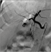
Right Hepatic Artery Pseudoaneurysm Following Laparoscopic Cholecystectomy: A Rare Complication Treated With Coil Embolization
- PMID: 40895958
- PMCID: PMC12398275
- DOI: 10.7759/cureus.89125
Right Hepatic Artery Pseudoaneurysm Following Laparoscopic Cholecystectomy: A Rare Complication Treated With Coil Embolization
Abstract
Hepatic artery pseudoaneurysm (HAPA) is an uncommon but potentially life-threatening vascular complication following laparoscopic cholecystectomy, often presenting days to weeks postoperatively. We describe the case of a 34-year-old female patient who presented 45 days following surgery with recurring hematemesis, melena, abdominal pain, and jaundice. Ultrasonography with colour Doppler suggested a vascular lesion near the porta hepatis, and triple-phase CT angiography confirmed a right hepatic artery (RHA) pseudoaneurysm leading to intrahepatic biliary radical dilatation (IHBRD). The patient underwent successful coil embolization, leading to full recovery without complications. HAPA should be considered in post-cholecystectomy patients who exhibit upper gastrointestinal (GI) hemorrhage or hemobilia. Early imaging diagnosis using Doppler ultrasound and CT angiography is imperative for timely intervention. Coil embolization provides a minimally invasive, effective alternative to open surgical repair, reducing associated morbidity and mortality. This case underscores the importance of prompt recognition and endovascular management of vascular complications following biliary surgery to prevent fatal outcomes.
Keywords: case report; coil embolization; hepatic artery injury; laparoscopic cholecystectomy; pseudoaneurysm; right hepatic artery.
Copyright © 2025, Babbar et al.
Conflict of interest statement
Human subjects: Informed consent for treatment and open access publication was obtained or waived by all participants in this study. Conflicts of interest: In compliance with the ICMJE uniform disclosure form, all authors declare the following: Payment/services info: All authors have declared that no financial support was received from any organization for the submitted work. Financial relationships: All authors have declared that they have no financial relationships at present or within the previous three years with any organizations that might have an interest in the submitted work. Other relationships: All authors have declared that there are no other relationships or activities that could appear to have influenced the submitted work.
Figures


Similar articles
-
Jaundice and bleeding caused by cystic artery pseudoaneurysm after cholecystectomy: A case report.Medicine (Baltimore). 2025 Aug 8;104(32):e43683. doi: 10.1097/MD.0000000000043683. Medicine (Baltimore). 2025. PMID: 40797483 Free PMC article.
-
Infective Endocarditis Following Coil Embolization for a Visceral Pseudoaneurysm: A Case Report.Cureus. 2025 Aug 6;17(8):e89484. doi: 10.7759/cureus.89484. eCollection 2025 Aug. Cureus. 2025. PMID: 40777070 Free PMC article.
-
Hemobilia as a result of right hepatic artery pseudoaneurysm rupture: An unusual complication of laparoscopic cholecystectomy.Int J Surg Case Rep. 2014;5(3):142-4. doi: 10.1016/j.ijscr.2014.01.005. Epub 2014 Jan 17. Int J Surg Case Rep. 2014. PMID: 24531018 Free PMC article.
-
Automated devices for identifying peripheral arterial disease in people with leg ulceration: an evidence synthesis and cost-effectiveness analysis.Health Technol Assess. 2024 Aug;28(37):1-158. doi: 10.3310/TWCG3912. Health Technol Assess. 2024. PMID: 39186036 Free PMC article.
-
Uterine artery embolization for symptomatic uterine fibroids.Cochrane Database Syst Rev. 2014 Dec 26;2014(12):CD005073. doi: 10.1002/14651858.CD005073.pub4. Cochrane Database Syst Rev. 2014. PMID: 25541260 Free PMC article.
References
-
- Radiological diagnosis and management of postlaparoscopic cholecystectomy right hepatic arterial pseudoaneurysm: a case report. Bhusal A, Jha SK, Oli R, Paudel B, Ghimire P. https://doi.org/10.1016/j.radcr.2024.09.005. Radiol Case Rep. 2024;19:6259–6264. - PMC - PubMed
-
- Newer anatomy of the liver and its variant blood supply and collateral circulation. Michels NA. https://doi.org/10.1016/0002-9610(66)90201-7. Am J Surg. 1966;112:337–347. - PubMed
-
- Surgical anatomy of the hepatic arteries in 1000 cases. Hiatt JR, Gabbay J, Busuttil RW. https://journals.lww.com/annalsofsurgery/abstract/1994/07000/surgical_an.... Ann Surg. 1994;220:50–52. - PMC - PubMed
-
- Right hepatic artery pseudoaneurysm post-laparoscopic cholecystectomy: a case report of endovascular stent-graft management. Ahmed S, Filep R, Mushtaq A, Budisca O. https://doi.org/10.7759/cureus.57127 Cureus. 2024;16:0. - PMC - PubMed
Publication types
LinkOut - more resources
Full Text Sources
