. 2025 Aug 18:30:629-640.
doi: 10.1016/j.reth.2025.08.003. eCollection 2025 Dec.

Conditioned plasma promotes full-thickness skin defect healing in a rat model

Affiliations

Conditioned plasma promotes full-thickness skin defect healing in a rat model

Majid Zamani et al. Regen Ther. .

Abstract

Introduction: Blood derivatives may enhance wound healing, but each possesses distinct characteristics and has yielded varying outcomes in patient treatment. This research seeks to examine the efficacy of conditioned plasma (CP) using polylactic acid (PLA) coated beads and to compare it with CP using bare beads and platelet-rich plasma (PRP) in the context of acute wound healing.

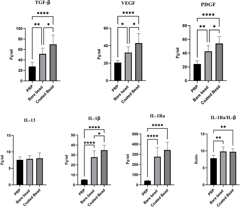
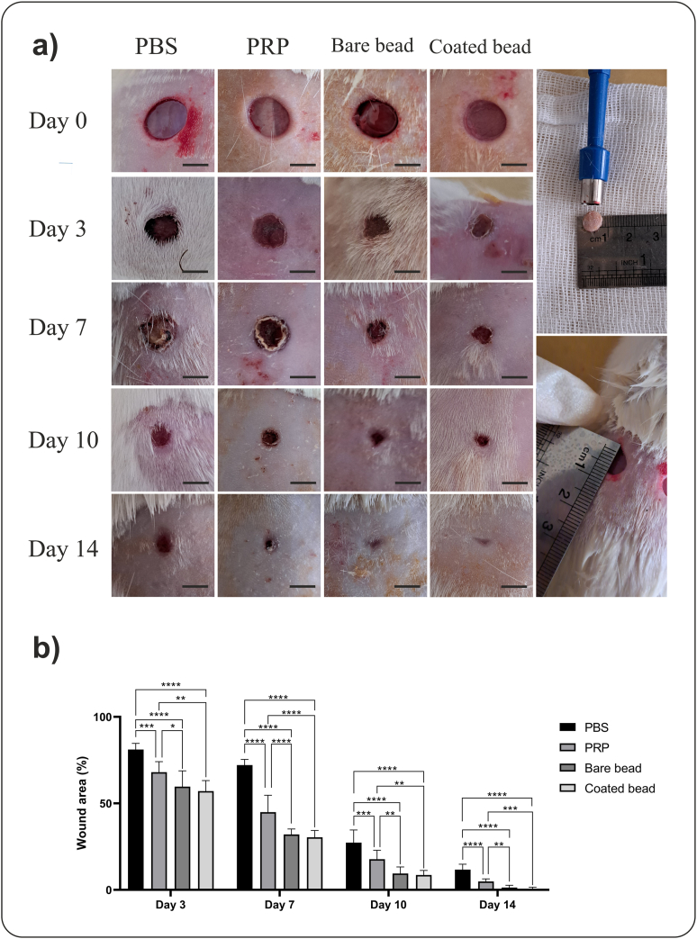
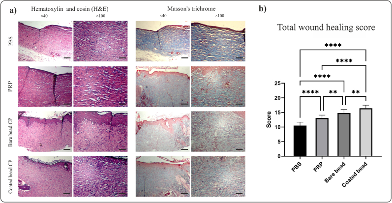
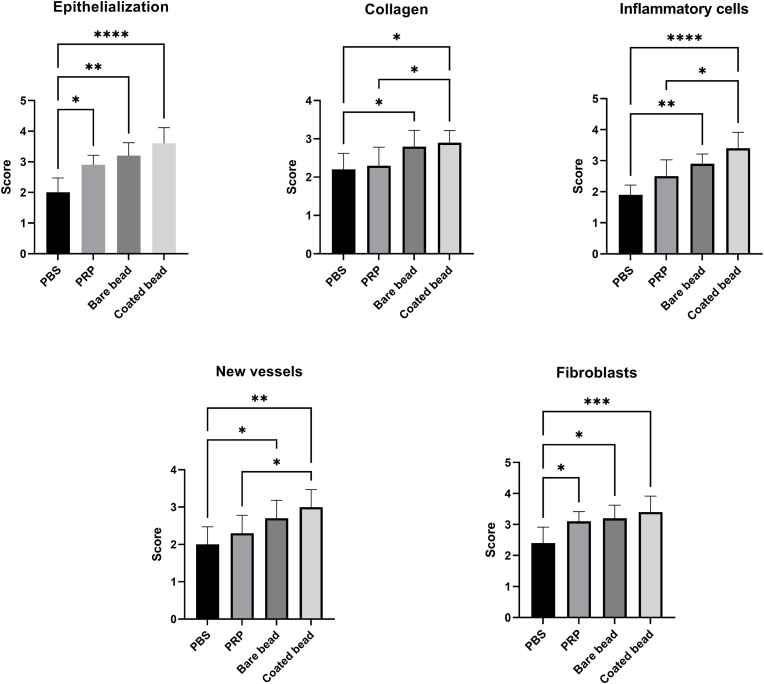
Methods: Blood was collected from 7 volunteer donors in three tubes containing ACD anticoagulant, PLA coated, or bare beads and incubated for 6 h at 37 °C. The concentration of VEGF, PDGF, TGF-β, IL-1β, IL-13, and IL-1Ra were measured by ELISA. Full-thickness wounds were made on the back of rats. PRP, CP with PLA-coated bead or bare beads, and phosphate buffer saline as control were administered to the wound area. Wound closure rate at days 3, 7, 10, and 14; epithelialization, fibroblast cells, inflammatory cells infiltration, new collagen formation, new vessel, and immunohistochemistry (CD31, α-SMA) were measured 14 days after the incision.

Results: The concentration of VEGF, PDGF, TGF-β, IL1-β, and IL-1Ra was significantly higher in CPs than in PRP (p < 0.05). CP with PLA-coated beads promoted wound closure and improved skin wound healing (p < 0.05), which was associated with enhanced epithelialization, fibroblast cell proliferation, new collagen formation, and reduced inflammatory cells infiltration. Immunohistochemistry showed an increase in CD31 and α-SMA levels in the treatment groups compared to the control group, but this increase was insignificant (p > 0.05).

Conclusion: CP promotes wound healing by increasing epithelialization, fibroblast proliferation, collagen synthesis and deposition, and reducing inflammatory cells infiltration.

Keywords: Conditioned plasma; Growth factor; Platelet rich plasma; Regenerative medicine; Wound healing.


Conflict of interest statement

Authors declare no conflict of Interests.

Figures

Image 1
Graphical abstract
Fig. 1
Fig. 1
Procedure for the preparation of blood derivatives. Blood was collected from seven healthy volunteers into tubes containing acid citrate dextrose (ACD) anticoagulant. For platelet-rich plasma (PRP) preparation, whole blood was centrifuged in a tube without beads to separate the plasma and buffy coat. The plasma was then centrifuged again to pellet the platelets, and the platelet-poor plasma (PPP) was discarded. The platelet pellet was resuspended in residual plasma to obtain PRP. For conditioned plasmas (CPs) preparation, whole blood was incubated in tubes containing either polylactic acid (PLA) coated beads or bare beads for 6 h at 37 °C and subsequently centrifuged to isolate the plasma. All derivatives were stored at −20 °C until further use.
Fig. 2
Fig. 2
Growth factors and cytokines concentration present in blood derivatives. The graph represents the vascular endothelial growth factor (VEGF), Platelet-derived growth factor (PDGF), transforming growth factor beta (TGF-β), Interleukin 13 (IL-13), Interleukin 1 beta (IL-1β), Interleukin 1 receptor antagonist (IL-1Ra), and ratio IL-1Ra/IL-1β levels in platelet rich plasma (PRP), conditioned plasma with bare beads (bare beads CP), and conditioned plasma with PLA coated beads (coated bead CP). Data are presented as mean ± SD of seven donors. (∗p < 0.05, ∗∗p < 0.01, ∗∗∗p < 0.001, ∗∗∗∗p < 0.0001).
Fig. 3
Fig. 3
Macroscopic analysis of wound healing. Conditioned plasma (CP) accelerates wound closure in a rat full-thickness excisional wound model. The rats' hair was shaved on different days to visualize the wounds better. a) Full-thickness wound images on days 0, 3, 7, 10, and 14 after wounding in the phosphate-buffered saline (PBS), platelet-rich plasma (PRP), bare beads CP, coated beads CP treatment groups, indicating better wound closure in the CPs groups (scale bars = 5 mm). The two images on the right depict the excisional wounds in rats immediately after creation (Day 0). b) The wound area percentage. The CPs treated group exhibited significantly accelerated wound closure compared to the other groups. Data are presented as mean ± SD of full-thickens wounds of 5 rats per group and two excisional wounds per rat (n = 10 wounds per group). Statistical analysis: ∗p < 0.05, ∗∗p < 0.01, ∗∗∗p < 0.01, ∗∗∗∗p < 0.0001.
Fig. 4
Fig. 4
Histological analysis of tissue sections at day 14 post wounding from full-thickness wounds in rat. Conditioned plasma (CP) accelerates wound healing in a rat full-thickness excisional wound model. a) Image of hematoxylin and eosin (H&E) and Masson's trichrome staining in different treatment groups, phosphate buffered saline (PBS), platelet-rich plasma (PRP), bare beads CP, coated beads CP (magnification × 40, scale bars = 200 μm; magnification × 100, scale bars = 100 μm). b) Total score from wound healing parameters. Total scores of wound healing indicating better wound healing in the group treated with coated beads CP. Data are presented as mean ± SD of full thickens wounds of 5 rats per group and two excisional wounds per rat (n = 10 wounds per group). Statistical analysis: ∗p < 0.05, ∗∗p < 0.01, ∗∗∗p < 0.01, ∗∗∗∗p < 0.0001.
Fig. 5
Fig. 5
Scoring of hematoxylin and eosin (H&E) and Masson's trichrome stained tissue sections. Various parameters were evaluated: epithelialization, inflammatory cells infiltration, new vessels, fibroblasts, and new collagen formation Data are presented as mean ± SD of full thickens wounds of 5 rats per group and two excisional wounds per rat (n = 10 wounds per group). Statistical analysis: ∗p < 0.05, ∗∗p < 0.01, ∗∗∗p < 0.01, ∗∗∗∗p < 0.0001.
Fig. 6
Fig. 6
Immunohistochemical staining of tissue sections at day 14 post wounding from full-thickness wounds in rat. a) Images of tissue sections of immunohistochemical staining for CD31 and α-SMA in different treatment groups, phosphate buffered saline (PBS), platelet-rich plasma (PRP), bare beads CP, coated beads CP (magnification × 40, scale bars = 200 μm; magnification × 100, scale bars = 100 μm). b) The modified Allred scoring system was used to score these two markers, which showed an increase in the CPs group compared to the other treatment groups, although this increase was not significant (p > 0.05).
Fig. 7
Fig. 7
Percentage of stained areas by immunohistochemical staining. The percentage quantification of CD31 and α-SMA positive area. The percentage of stained areas was higher in the CPs treated groups, although, this difference was not significant (p > 0.05). Data are presented as mean ± SD of full thickens wounds of 5 rats per group and two excisional wounds per rat (n = 10 wounds per group).

Similar articles

References

    1. Nouri S., Shokraneh S., Fatehi Shalamzari P., Ahmed M.H., Radi U.K., Idan A.H., et al. Application of mesenchymal stem cells and exosome alone or combination therapy as a treatment strategy for wound healing. Cell Biochem Biophys. 2024;82:3209–3222. doi: 10.1007/s12013-024-01448-w. - DOI - PubMed
    1. Sorg H., Sorg C.G.G. Skin wound healing: of players, patterns, and processes. Eur Surg Res. 2022;64:141–157. doi: 10.1159/000528271. - DOI - PubMed
    1. desJardins-Park H.E., Gurtner G.C., Wan D.C., Longaker M.T. From chronic wounds to scarring: the growing health care burden of under- and over-healing wounds. Adv Wound Care. 2022;11:496–510. doi: 10.1089/wound.2021.0039. - DOI - PMC - PubMed
    1. Daneste H., Mohammadzadeh Boukani L., Ramezani N., Asadi F., Zaidan H.K., Sadeghzade A., et al. Combination therapy along with mesenchymal stem cells in wound healing; the state of the art. Adv Med Sci. 2023;68:441–449. doi: 10.1016/j.advms.2023.10.006. - DOI - PubMed
    1. Rodrigues M., Kosaric N., Bonham C.A., Gurtner G.C. Wound healing: a cellular perspective. Physiol Rev. 2019;99:665–706. doi: 10.1152/physrev.00067.2017. - DOI - PMC - PubMed

LinkOut - more resources