. 2025 Sep;292(2054):20250489.
doi: 10.1098/rspb.2025.0489. Epub 2025 Sep 3.

Cellular, bone-like tissue in the bucklers and thorns of the thornback ray Raja clavata (Batoidea, Chondrichthyes)

Affiliations

Cellular, bone-like tissue in the bucklers and thorns of the thornback ray Raja clavata (Batoidea, Chondrichthyes)

Aaron R H LeBlanc et al. Proc Biol Sci. 2025 Sep.

Abstract

Chondrichthyans (cartilaginous fishes) have lost the cellular bone characteristic of other jawed vertebrate skeletons. However, we identify cellular bone-like tissue in modified scales with enlarged bases, called 'bucklers' and 'thorns', which are distinctive for one group of extant batoids (rays). As placoid scales, they possess spines of orthodentine and osteodentine, but a unique basal structure. This consists of a cell-rich material, previously misidentified as an acellular tissue. Newly formed basal tissue grows appositionally and episodically from a cell-rich periosteum-like layer and closely resembles cellular bone, with entombed cells situated between bundles of attachment fibres anchoring the scale to the underlying dermal tissue and the 'periosteum' to the scale surface. In histologically more mature tissue, the cell spaces and attachment fibres are remodelled, forming enlarged, elongated spaces. The result is a unique mineralized tissue in these rays, initially sharing similarities with cellular bone, but with a mature state where cell spaces are modified throughout the base, by proposed remodelling of the matrix. Our findings of cellular bone forming the attachment tissues in ray scales demonstrate the chondrichthyan capacity to deposit bone-like tissues within the odontode module, contrary to previous understandings of hard tissue evolution in vertebrates.

Keywords: Batoidea; Chondrichthyes; Raja clavata; bone; bucklers; placoid scales; skin denticles; thorns.


Conflict of interest statement

We declare a competing interest. The lead author was an associate editor for Proceedings B at the time of submission of this article.

Figures
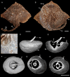
Distribution of thorns and bucklers in extant female Raja clavata.
Figure 1.
Distribution of thorns and bucklers in extant female Raja clavata (Rajidae, Chondrichthyes) and the gross external and internal anatomy of a buckler. A, BMNH 64.4.26:87-8, smaller individual dominated by symmetrically positioned bucklers; B, BMNH 85.11.3.4, larger, putatively older individual with more bucklers present, more thorns have been added (on the tail, fin edges). C, Ventral view of a freshly caught R. clavata showing numerous bucklers around the jaws, gill openings and farther posteriorly (specimen was released after being photographed). D, Lateral view of a CT scanned buckler (BMNH 2017.5.12.9, NHM Life Sciences collection) showing the expanded base. E, Virtual longitudinal section. F, Dorsal view of the buckler. G, Virtual horizontal section through the buckler near the dorsal surface of the basal tissue. H, Virtual horizontal section farther ventrally than in G. Scale bars in A, B = 5 cm, scale bars in D–H = 0.5 cm. ba, buckler base; bu, buckler; bu.sp, buckler near spiracle; dep, posterior depression of buckler spine; go, gill opening; j, jaws; pc, pulp cavity; rl, rest line; sp, spine of buckler spine; th, thorn; U, ‘U’-shaped bend along anterior margin of the spine. White asterisk indicates the position of posterior overgrowth of tissue impinging upon the spine.
Hard tissue histology of the major tissue types in a buckler of Raja clavata.
Figure 2.
Hard tissue histology of the major tissue types in a buckler of Raja clavata (Rajidae, Chondrichthyes, unregistered specimen, NHM Life Sciences collection). A–N, sections through buckler shown in figure 1. A, C–F, H, I, L, plane-polarized light; B, G, J, K, M, N, cross-polarized light. A, Longitudinal serial section, letters B–N indicate regions in close-up on the remainder of the plate, black arrow indicates basal layer shown in more detail in figure 5. Black arrowheads indicate growth lines; B, point where the spine meets the buckler base, dominated by Sharpey’s fibres; C, tip of the buckler spine, asterisk indicates wear at the tip; D, posterior margin of the spine (equivalent to region marked by asterisks in figure 1 D–F); E, base of the spine with ortho- and osteodentine, buckler base below with microcancellous tissue; F, G, base of the spine with orthodentine and microcancellous tissue below; H, K, microcancellous tissue with denser spaces; I, J, microcancellous tissue with less dense spaces, intrinsic fibres (asterisk) apparent in cross-polarized light (J); L–N, microcancellous tissue with dense intrinsic fibres distributed throughout; N, close-up of region in M, to show scalloped surfaces indicative of osteoclast-like activity and resorption (arrowheads). de, denteon; enam, enameloid; mic, microcancellous tissue; ord, orthodentine; osd, osteodentine; sh.f, Sharpey’s fibres; tub, dentine tubules; numbers 1−4 indicate equivalent microcancellous tissue spaces in F, G, H, K, I, J, L–N. Scale bars: A = 1000 µm, B–G, I, H = 25 µm.
Hard tissue histology of the basal tissues in a buckler of Raja clavata.
Figure 3.
Hard tissue histology of the basal tissues in a buckler of Raja clavata (Rajidae, Chondrichthyes, unregistered specimen, NHM Life Sciences collection). A–G, H–K, longitudinal sections in A, C, F, H, white light; B, D, E, G, I, K, cross-polarized light. J, Image of sectioned buckler in dorsal view. A, B, Similar to buckler in figure 5, but with a thicker region of osteodentine (denteon shown in inset A from the osteodentine) and multiple layers of orthodentine in the spine. Asterisk indicates the region where the close-up image of denteons in the osteodentine layer was taken; in A, letters C–I indicate regions shown in close-up in the rest of the figure. C, D, Spine base (orthodentine) and osteodentine, in cross-polarized light; (D) denteons in the osteodentine surrounded by fibres organized in a Maltese cross pattern; this is less apparent further along the spine base (E); F, G, transition from the osteodentine to the microcancellous tissue, fibre arrangement is lacking in cross-polarized light (G); H, I, K, base of the buckler with a distinct outer layer, rich in intrinsic fibres that parallel the buckler base (asterisk). Small spaces are present within this fibre layer (1–3), surrounded by the fibres. White arrowheads indicate large spaces with the microcancellous tissue (H, I), with potential evidence of tissue resorption (K, white arrowheads). de, denteon; dep, depression; enam, enameloid; mic, microcancellous tissue; ord, orthodentine; osd, osteodentine; pc, pulp cavity; sh.f, Sharpey’s fibres; tub, dentine tubules; numbers 1−5 indicate equivalent spaces within the osteodentine (C, D, F, G) and in the basal tissue (H, I). Scale bars: A = 1 mm; C–I = 100 µm; J, K = 0.5 cm.
Small buckler of Raja clavata.
Figure 4.
Small buckler of Raja clavata (Rajidae, Chondrichthyes, unregistered specimen, NHM Life Sciences collection) with a less expanded base. A, Longitudinal section under plane-polarized light of the buckler shown in dorsal view in the inset. B, Longitudinal section in cross-polarized light. C, D, Region of microcancellous tissue with rounded spaces and showing a Maltese cross arrangement of fibres around the spaces (D). E, Close-up of microcancellous tissue showing spaces with irregular walls, reminiscent of Howship’s lacunae. F, Close-up of a second region with putative Howship’s lacunae around the enlarged spaces. G, Cross-polarized light close-up image of microcancellous tissue showing putative Howship’s lacunae. enam, enameloid; Hl?, putative Howship’s lacunae; mic, microcancellous tissue; mt, matrix; ord, orthodentine; pc, pulp cavity; sp, spaces in microcancellous tissue; numbers 1−5 indicate equivalent spaces within the basal tissue (C, D). Scale bars: A, B = 1 mm; C, D = 100 µm.
Soft tissue histology of bucklers from a Raja clavata.
Figure 5.
Paraffin histology of bucklers from a Raja clavata (Rajidae, Chondrichthyes), France. A, C, D–F, Decalcified, stained longitudinal sections; B, macrophotograph of Raja pectoral fin with in situ bucklers. A, Buckler section with broken spine (PAS-BA stain), white asterisks indicate regions of buckler base growing over the fin musculature; C, dorsal region of buckler (Hematoxylin & Eosin stain) showing three distinct regions (mic, mat, fib) indicating different stages of buckler development. Black arrowheads indicate single cells within the microcancellous spaces. Circled region shows a cell-free region of large spaces and sparse matrix; D, second stained buckler (Masson’s trichrome) section; E, close-up of the interface between the three major regions of the buckler base in C. Black asterisk indicates matrix-producing cells being engulfed by matrix (along with thicker fibres, entombed cells are aligned between these fibres). ‘+’ Indicates angular cell spaces, potentially indicating osteocytic osteolysis; F, close-up of ventral surface of buckler in D showing regions of peripheral bone-like layer with osteocytes and more internal layers of maturing microcancellous tissue with enlarged cell lacunae, suggestive of osteocytic osteolysis (+); G, section comparing blood vessel spaces in the buckler to the microcancellous spaces; H, transect through the buckler base in A (but with H&E stain), showing histological interpretations of the major tissue types forming the base. Black arrowheads identify cells within the microcancellous tissue. White arrowheads mark potential transitional cells with enlarged lacunae, interpreted as mononucleated cells. bt, bone-like tissue; bv, blood vessel; der, dermis; fib, fibrous layer; mat, mineralized matrix; mic, microcancellous tissue; ord, orthodentine; osd, osteodentine; osl, osteocyte lacunae; po, periosteum-like layer; rl, rest line; sh.f, Sharpey’s fibres; sp, spaces in microcancellous tissue.
Hypothetical growth series of bucklers and thorns, schematic illustration of young and mature bucklers, highlighting the growth of the basal tissues.
Figure 6.
Hypothetical growth series of bucklers and thorns, schematic illustration of young and mature bucklers, highlighting the growth of the basal tissues. A, Small scale (buckler or thorn) with incipiently developed basal tissue and an open pulp cavity in longitudinal section. B, Slightly larger scale with more extensively developed basal tissues and a partially enclosed pulp. C, Larger scale with a nearly completely enclosed pulp and thicker mass of basal tissue. D, Close-up of periphery of basal tissue in A, showing outer bone-like and inner microcancellous tissues. E, Close-up of periphery of basal tissue in B, showing outer bone-like and inner microcancellous tissues. F, Close-up of periphery of basal tissue in C, showing outer bone-like and inner microcancellous tissues separated by multiple rest lines. G, Illustration of a young scale with interpretation of basal tissues and their growth directions. H, Overview illustration of a mature buckler. The basal tissue is formed of successive layers of mature, microcancellous tissue (light pink), which is derived from the same cellular bone-like tissue forming along the periphery (dark pink) in the youngest scales (G). I, Illustration of the formation and maturation of the basal buckler tissues, highlighting the formation of the cellular bone-like tissue from the periosteum-like layer (red) and the distribution of the three cell types (‘osteoblast’, ‘osteocyte’ and mature cells). bo, cellular bone-like layer; cl, cell lacunae; de, dentine; enam, enameloid; gl, growth line; mic, microcancellous tissue; ord, orthodentine; osd, osteodentine; po, periosteum-like layer; pu, pulp; sh.f, Sharpey’s fibres.

Similar articles

References

    1. Reif W. 1974. Morphogenese und Musterbildung des Hautzähnchen‐Skelettes von Heterodontus. Lethaia 7, 25–42. ( 10.1111/j.1502-3931.1974.tb00882.x) - DOI
    1. Reif WE. 1978. Protective and hydrodynamic function of the dermal skeleton of elasmobranchs. Neues Jahr. Geol. Paläontol. Abhand. Paleoecol. 157, 133–141.
    1. Reif WE. 1985. Squamation and ecology of sharks. Cour Forsch. Senckenberg 78, 1–255.
    1. Oeffner J, Lauder GV. 2012. The hydrodynamic function of shark skin and two biomimetic applications. J. Exp. Biol. 215, 785–795. ( 10.1242/jeb.063040) - DOI - PubMed
    1. Wen L, Weaver JC, Thornycroft PJM, Lauder GV. 2015. Hydrodynamic function of biomimetic shark skin: effect of denticle pattern and spacing. Bioinspir. Biomim. 10, 066010. ( 10.1088/1748-3190/10/6/066010) - DOI - PubMed

MeSH terms

LinkOut - more resources