. 2025 Sep 2;16(1):668.
doi: 10.1038/s41419-025-07988-0.

Tissue-specific iron levels modulate lipid peroxidation and the FLASH radiotherapy effect

Affiliations

Tissue-specific iron levels modulate lipid peroxidation and the FLASH radiotherapy effect

Nuria Vilaplana-Lopera et al. Cell Death Dis. .

Abstract

Iron is vital to living cells, playing a key role in cellular respiration, DNA synthesis, and various metabolic functions. Importantly, cancer cells have a higher dependency on iron compared to normal cells to support their rapid growth and survival. Due to this fact, tumors are more vulnerable to ferroptosis, an iron-dependent form of regulated cell death. Radiation therapy (RT), a standard treatment for many cancer patients, is known to induce ferroptosis. Ultra-high dose rate FLASH RT offers an improved therapeutic window by minimizing damage to normal tissues while preserving tumor control. However, the precise biological mechanisms behind the protective effects of FLASH RT on normal tissues remain unclear. In this study, we propose that variations in lipid peroxidation and ferroptosis, driven by intrinsic differences in iron levels between normal and cancerous tissues, contribute to this effect. Our findings show that FLASH RT increases lipid peroxidation and induces ferroptosis in tumor cells but does not significantly elevate lipid peroxidation and ferroptosis in normal tissues compared to conventional RT. To determine whether raising iron levels in normal tissues could abrogate the protective effects of FLASH, mice were fed a high-iron diet before RT. A high-iron diet before and after RT reversed the protective effect of FLASH, resulting in increased intestinal damage and lipid peroxidation. This suggests that baseline iron levels and iron-driven lipid peroxidation are critical factors in mediating the protective outcomes of FLASH RT. Overall, our study sheds light on the role of iron in modulating RT responses and provides new mechanistic insights into how FLASH RT influences normal and cancerous tissues.


Conflict of interest statement

Competing interests: The authors declare no competing interests.

Figures
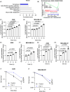
Fig. 1
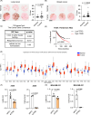
Fig. 1. RT induces lipid peroxidation and ferroptosis in cancer cells in a dose and time-dependent manner.
A RNA sequencing was performed after treating MDA-MB-231 cells with 0 or 10 Gy of RT. Pathways analysis by Enrichr identified that genes involved in the “Cell Cycle”, “p53 signaling pathway”, “DNA replication” and “Ferroptosis” were significantly altered by RT. B GSEA enrichment further confirmed significant alterations of genes involved in ferroptosis after RT. Lipid peroxidation was measured using C11 BODIPY in A549 and MDA-MB-231 24 h after RT at varying doses from 2 to 10 Gy (C, D) as well as at different time points after 10 Gy of RT (EH). Clonogenic survival was determined after RT with or without pre-treatment of 20 µM (A549, I) or 4 µM (MDA-MB-231, J) Ferr-1. RT was given using a 137Cs irradiator. Error bars indicate standard deviation (SD) (n = 3 per group). Statistical tests were performed by One-way ANOVA with Dunnett’s multiple comparisons test (CH) and unpaired t-test (I, J).
Fig. 2
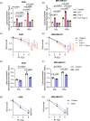
Fig. 2. FLASH RT induces similar levels of lipid peroxidation and ferroptosis with conventional RT in cancer cells.
A, B A549 (A) and MDA-MB-231 (B) cells were irradiated at 10 Gy with conventional or FLASH dose rates with an electron linear accelerator. A significant increase in lipid peroxidation was observed 24 h after RT using C11 BODIPY in all treatment groups. C, D A significant dose-dependent induction of lipid peroxidation was observed after conventional and FLASH RT. E, F Clonogenic survival was determined after conventional or FLASH RT with or without pre-treatment of 20 µM (A549) or 4 µM (MDA-MB-231) Ferr-1. Error bars indicate standard deviation (SD) (n = 3 per group). Statistical tests were performed by One-way ANOVA with Dunnett’s multiple comparisons test (A, B), Two-way ANOVA with Tukey’s multiple comparisons test (C, D), and One-way ANOVA with Tukey’s multiple comparisons test (E, F).
Fig. 3
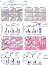
Fig. 3. FLASH RT induces lipid peroxidation in mouse xenograft tumors while sparing normal lung and intestine tissues.
A Lung tumor tissues (A549 and Calu-6) from subcutaneous mouse xenograft models were stained with 4-HNE, a lipid peroxidation marker. After conventional and FLASH RT (15 Gy for A549 and 20 Gy for Calu-6), there was a significant increase in 4-HNE staining intensity (n = 4 for A549 0 Gy and conventional RT, n = 3 for FLASH RT, n = 6 for Calu-6 0 Gy and FLASH RT, n = 8 for Calu-6 conventional RT). B BALB/c mouse lung tissues were stained with 4-HNE after 10 Gy of conventional or FLASH RT. Lipid peroxidation was significantly enhanced 24 h and 7 days after conventional RT. However, FLASH RT did not change lipid peroxidation (n = 6 per group at each time point). C, D BALB/c mice were treated daily with 2 mg/kg Ferr-1 or DMSO vehicle, starting one day prior to RT, to inhibit ferroptosis. Lipid peroxidation, detected by 4-HNE staining, was markedly increased after 10 Gy conventional RT compared to 0 Gy, but not after 10 Gy FLASH RT in the DMSO-treated group (C). This increase in lipid peroxidation was reversed by Ferr-1 treatment. Tissue damage in the upper intestines was assessed by H&E staining, based on the number of remaining intestinal crypts (D, arrowheads). Both 10 Gy conventional and FLASH RT significantly reduced crypt numbers, with conventional RT causing more severe damage. Ferr-1 treatment improved crypt preservation in the conventional RT group but had no significant effect in the 0 Gy or FLASH RT groups (n = 4/group). Error bars indicate standard deviation (SD). Statistical tests were performed by One-way ANOVA (A) or Two-way ANOVA (BD) with Tukey’s multiple comparisons test. IntD: Integrated Density.
Fig. 4
Fig. 4. Iron is essential for tumor survival.
A, B Tissue microarray slides from breast and lung cancer patients were analyzed to measure iron levels using Prussian blue. Compared to normal lung (A) or breast tissues (B), iron levels were significantly higher in lung and breast cancer tissues (Lung cancer; n = 20 for normal tissues, n = 32 for lung adenocarcinoma tissues, Breast cancer; n = 11 for normal tissues, n = 90 for cancer tissues). C The functional enrichment analysis of 748 genes from the Catalogue of Somatic Mutations in Cancer (COSMIC) Cancer Gene Consensus showed significantly regulated pathways of cancer driving genes “4 iron 4 sulfur cluster binding”, “response to iron ion”, and “cellular iron ion binding”. D Analysis of TCGA PanCancer Atlas data revealed that cancer patients exhibiting higher TFRC expression in tumor tissues compared to normal tissues had significantly poorer overall survival outcomes (Low TFRC: n = 2014, High TFRC: n = 3257; log-rank test, p < 0.0001). E TCGA data showing TFRC mRNA expression in normal and various cancer patient tissues were visualized using UALCAN. F, G The role of iron in tumor cell survival was determined by knocking down TFRC, an iron transporter. Compared to the siRNA against scrambled sequence, the inhibition of TFRC expression significantly decreased intracellular iron and cell survival measured by clonogenic assay in both A549 (F) and MDA-MB-231 (G) cells. Statistical tests were performed by nonparametric Kolmogorov–Smirnov test (A, B), log-rank (C), and upaired t-test (F, G).
Fig. 5
Fig. 5. Iron enhances RT sensitivity and ferroptosis.
AD The role of iron in lipid peroxidation and cell survival was determined by measuring lipid peroxidation using C11 BODIPY and clonogenic assay, respectively. Adding a suboptimal dose of iron (50 μM and 5 μM of ammonium iron (II) sulfate in A549 and MDA-MB-231 respectively) increased lipid peroxidation (A, B) and radiosensitization (C, D) in both cell lines. Treatment with Ferr-1 (20 μM and 4 μM in A549 and MDA-MB-231, respectively) decreased lipid peroxidation induced by RT (A, B) and reversed radiosensitization (C, D) indicating that ferroptosis was further induced by external iron. EH Inhibition of iron availability using deferoxamine (DFO) (200 nM and 100 nM in A549 and MDA-MB-231, respectively) decreased lipid peroxidation induced by RT (E, F) and increased radioresistance (G, H) of A549 and MDA-MB-231 cells. (AH, n = 3/group) Error bars indicate standard deviation (SD). Two-way ANOVA with Tukey’s multiple comparisons test (A, B), One-way ANOVA with Tukey’s multiple comparisons test (C, D, G, H), and unpaired t-test (E, F). For (C, D, G, H), 6 Gy conditions were compared.
Fig. 6
Fig. 6. Increasing iron levels in normal tissue reverts FLASH sparing effect.
A BALB/c mice were fed with high iron diet (5000 ppm iron) for 24 h to increase iron levels in normal intestine. After RT treatment at 8 or 10 Gy, mice were returned to a normal diet or stayed on a high-iron diet for another 72 h. (HI: High iron diet). B, C Lipid peroxidation, which was stained with 4-HNE was highly increased by both 8 (B) and 10 Gy (C) conventional RT in the control or high iron diet groups both for 24 h and 96 h. FLASH RT at 8 or 10 Gy did not increase lipid peroxidation as significantly as conventional RT with the control or 24-h high-iron diet. However, a prolonged high-iron diet even after RT treatment eliminated the FLASH effect on lipid peroxidation. D, E Tissue damage in the upper intestines was determined by H&E staining. The presence of remaining intestinal crypts (arrow heads) was counted and analyzed. The number of remaining crypts was significantly decreased after 8 (D) and 10 Gy of conventional RT in the control or high-iron diet groups for 24 h and 96 h. FLASH RT-induced damages were significantly lower than conventional RT in the control or 24-h high-iron diet mice. However, a prolonged high iron diet, diminished the sparing observed in FLASH irradiated tissues since FLASH RT induced similar tissue damage to conventional RT. (BE, for 8 Gy, n = 8 in every condition; for 10 Gy, control diet n = 12, HI diet 24 h or 96 h n = 6) Error bars indicate standard deviation (SD). Statistical test was performed by Two-way ANOVA with Tukey’s multiple comparisons. IntD Integrated Density.

Update of

References

    1. Federico G, Carrillo F, Dapporto F, Chiariello M, Santoro M, Bellelli R, et al. NCOA4 links iron bioavailability to DNA metabolism. Cell Rep. 2022;40:111207. - PubMed
    1. Torti SV, Manz DH, Paul BT, Blanchette-Farra N, Torti FM. Iron and cancer. Annu Rev Nutr. 2018;38:97–125. - PMC - PubMed
    1. Hann HW, Stahlhut MW, Menduke H. Iron enhances tumor growth. Observation on spontaneous mammary tumors in mice. Cancer. 1991;68:2407–10. - PubMed
    1. Radulescu S, Brookes MJ, Salgueiro P, Ridgway RA, McGhee E, Anderson K, et al. Luminal iron levels govern intestinal tumorigenesis after Apc loss in vivo. Cell Rep. 2012;2:270–82. - PubMed
    1. Sukiennicki GM, Marciniak W, Muszynska M, Baszuk P, Gupta S, Bialkowska K, et al. Iron levels, genes involved in iron metabolism and antioxidative processes and lung cancer incidence. PLoS ONE. 2019;14:e0208610. - PMC - PubMed

LinkOut - more resources