

Published Erratum
doi: 10.1038/s41598-025-13503-4.
Author Correction: hsa_circ_0008305 facilitates the malignant progression of hepatocellular carcinoma by regulating AKR1C3 expression and sponging miR-379-5p
Affiliations
- PMID: 40897744
- PMCID: PMC12405477
- DOI: 10.1038/s41598-025-13503-4
Item in Clipboard
Published Erratum
Author Correction: hsa_circ_0008305 facilitates the malignant progression of hepatocellular carcinoma by regulating AKR1C3 expression and sponging miR-379-5p
Sci Rep.
.
No abstract available
Figures
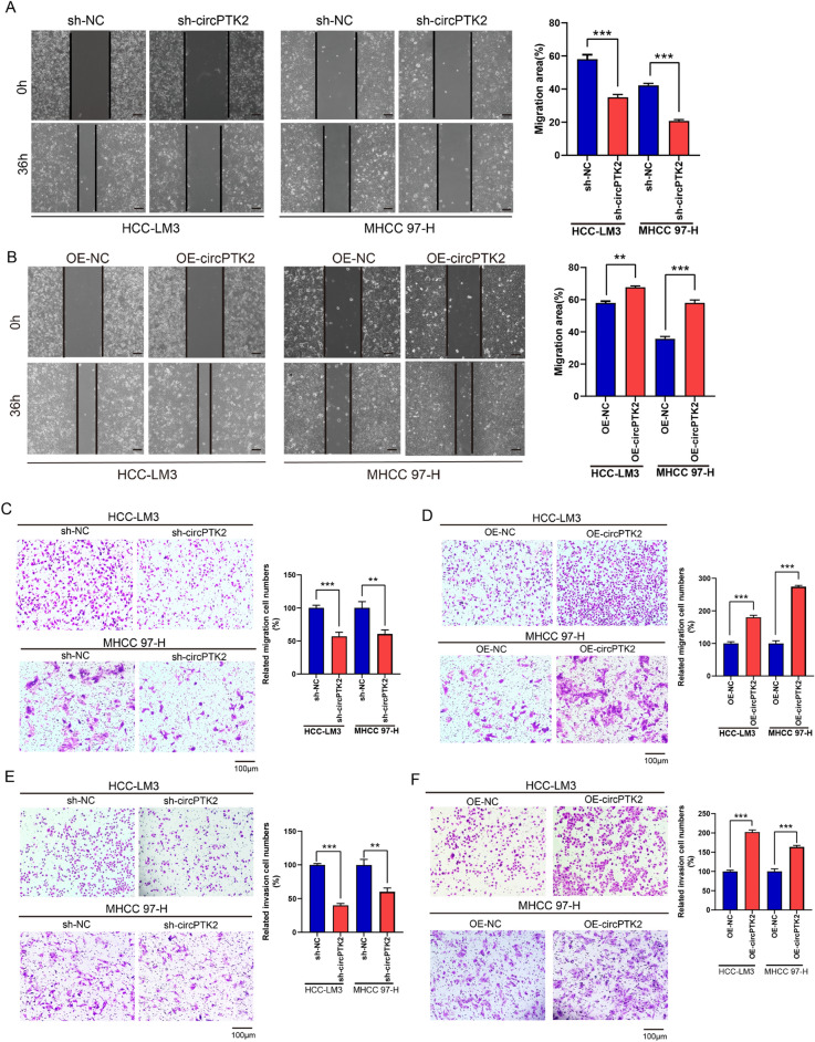
circPTK2 increases the migration and invasion capability of HCC cells. (A,B) Cell migration ability of HCC-LM3 and MHCC 97-H cells transfected with sh-circPTK2 plasmid(A), or OE-circPTK2 plasmid(B), assessed by scratch-healing assays; Mean ± SD, n = 3, **p < 0.01, ***p < 0.001, scale bar:100 μm. (C,D) The transwell assays without Matrigel evaluate the migration ability of HCC-LM3 and MHCC 97-H after circPTK2 knockdown(C) or overexpressing(D); Mean ± SD, n = 3, **p < 0.01, ***p < 0.001. (E,F) The invasion capability of HCC cells transfected with sh-circPTK2 plasmid(E), or OE-circPTK2 plasmid(F), was examined using transwell assays with Matrigel; Mean ± SD, n = 3, **p < 0.01, ***p < 0.001, vs. corresponding control plasmids.
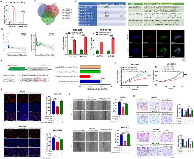
Identification of miRNA binding to circPTK2 that regulates the proliferation, migration and invasion in HCC cells. (A) RIP assays validated the combination of circPTK2 with anti-AGO2 in HCC-LM3 and MHCC 97-H cells (Mean ± SD, n = 3, ***p < 0.001). (B) Venn plot showed the miRNAs combination to AKR1C3 predicted by miRDB, Targetscan, and mirDIP. (C) miRNAs combination sites with circPTK2 forecast by miRanda v3.3a. (D) Spearman correlation analyses showing the relationships of miR-379-5p and AKR1C3 (left), and circPTK2 (right) in HCC tissues (n = 72). (E) Biotin-labeled miRNA pull-down assay confirmed the interaction of miR-379-5p with circPTK2 and AKR1C3 (Mean ± SD, n = 3, ***p < 0.001). (F) The co-location of miR-379-5p and circPTK2 in HCC cells judged by FISH. (G) Luciferase reporter assay detected the luciferase activity in HEK-293T cells co-transfected with miRNA-379-5p mimic (or mimic-NC) and circPTK2-wt (or circPTK2-mut) luciferase reporter vectors. Mean ± SD, n = 3, ***p < 0.001. (H) Cell viability of HCC-LM3 and MHCC 97-H cells transfected with sh-circPTK2 alone or co-transfected with miR-379-5p inhibitor, measured by CCK8(Mean ± SD, n = 6, ***p < 0.001). (I) EdU assays estimates the DNA replication capacity of HCC-LM3 and MHCC 97-H cells transfected with sh-circPTK2 alone or co-transfected with miR-379-5p inhibitor. Mean ± SD, n = 3, **p < 0.01, ***p < 0.001, scale bar: 100 μm. (J,K) The cells migration of HCC-LM3 and MHCC 97-H co-transfected with sh-circPTK2 and miR-379-5p inhibitor, assessed by scratch-healing experiments (J) and transwell migration assays (K). Mean ± SD, n = 3, ***p < 0.001, scale bar:100 μm. (L) The invasion ability of HCC-LM3 and MHCC 97-H transfected with sh-circPTK2 and miR-379-5p inhibitor, measured by transwell invasion assays. Mean ± SD, n = 3, ***p < 0.001.
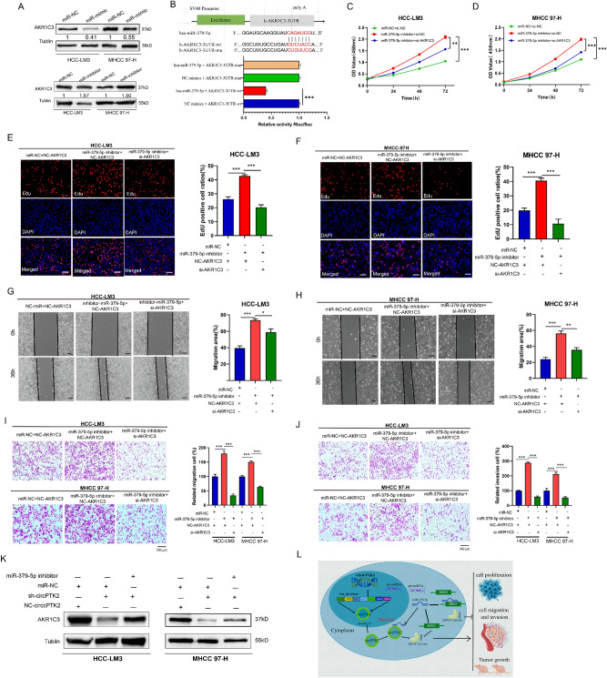
miR-379-5p targets AKR1C3 to sustain HCC progression. (A) Western blot detected the protein expression of AKR1C3 in HCC-LM3 and MHCC 97-H transfected with miR-379-5p mimic or inhibitor.(B) Schematic of AKR1C3 3′ UTR wild-type (wt) and mutant (mut) luciferase reporter vectors; Luciferase activity of HEK-293T cell co-transfected with miR-379-5p mimic and AKR1C3 3′-UTR (wt) or AKR1C3 3′-UTR (mut) vectores determined by dual-luciferase reporter assays, Mean ± SD, n = 3, ***p < 0.001. (C,D) Cell viability of HCC cells transfected with miR-379-5p inhibitor alone or co-transfected with si-AKR1C3, measured by CCK8 (Mean ± SD, n = 6, **p < 0.01, ***p < 0.001). (E,F) DNA replication capacity of HCC-LM3 (E) and MHCC 97-H (F) cells co-transfected with miR-379-5p inhibitor and si-AKR1C3, evaluated by EdU assays (Mean ± SD, n = 3, ***p < 0.001). scale bar: 100 μm. (G–I) The cells migration of HCC-LM3 and MHCC 97-H transfected with miR-379-5p inhibitor and si-AKR1C3, assessed by scratch-healing experiments (G,H) and transwell migration assays (I). Mean ± SD, n = 3, *p < 0.05, **p < 0.01, ***p < 0.001, scale bar:100 μm. (J). The invasion ability of HCC cells transfected with miR-379-5p inhibitor and si-AKR1C3, measured by transwell invasion assays. Mean ± SD, n = 3, ***p < 0.001, scale bar:100 μm. (K) Western blot analyzed the expression of AKR1C3 in HCC-LM3 and MHCC 97-H co-transfected with sh-circPTK2 and miR-379-5p inhibitor. (L) Schematic diagram shows circPTK2 promoting HCC cell proliferation, migration and invasion via miR-379-5p/AKR1C3 axis.
Erratum for
-
hsa_circ_0008305 facilitates the malignant progression of hepatocellular carcinoma by regulating AKR1C3 expression and sponging miR-379-5p.Sci Rep. 2025 Jan 8;15(1):1309. doi: 10.1038/s41598-025-85737-1. Sci Rep. 2025. PMID: 39779892 Free PMC article.
Publication types
LinkOut - more resources
Full Text Sources
