. 2025 Sep 2;15(1):32291.
doi: 10.1038/s41598-025-17336-z.

Establishment of Drosophila intestinal cell lines as tools for multiomic screening and deciphering intestinal biology

Affiliations

Establishment of Drosophila intestinal cell lines as tools for multiomic screening and deciphering intestinal biology

Arthur Luhur et al. Sci Rep. .

Abstract

The Drosophila intestine is a powerful model for stem cell dynamics and epithelial biology, yet no intestinal derived cell lines have been available until now. Here, we describe the establishment of Drosophila intestinal cell lines. The cell lines were derived from the embryonic intestine through specific RasV12 expression and show continuous proliferation and the ability to be frozen and re-thawed. Each derived cell line exhibited morphological and cellular heterogeneity. Single-cell RNA sequencing confirmed their intestinal origin and cell populations with unique enriched signaling pathways. In addition, L15, one of the three lines, formed 3D spheroids that displayed epithelial polarity. Together, these lines provide an additional resource for studying intestinal development, epithelial organization, and pest management.

Keywords: Drosophila; Cell line; Intestine; Spheroids; Transcriptomics.


Conflict of interest statement

Declarations. Competing interests: The authors declare no competing interests.

Figures

Fig. 1
Fig. 1
CG10116-GAL4 drives intestinal-specific GFP expression in embryos and adults. Micrographs of Stage 13 to Stage 17 wildtype (A) or RasV12 expressing (B) embryos. To the left of each panel are merged images with DAPI and GFP (yellow) and the images to the right are GFP channel only. Micrographs of the GFP channel highlighting intestinal morphology of the embryos expressing RasV12 in the intestinal cells are displayed to the right. GFP expression driven by CG10116-GAL4 was also characterized in either control (C) or RasV12 (D) adult male guts. The respective brightfield (C’,D’) and GFP channel (C’’, D’’) are also shown. Scale bars = 100 μm.
Fig. 2
Fig. 2
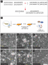
Strategy to create and morphology of embryonic intestine derived cell lines. (A)The schematic for the genetic cross to generate embryos carrying the CG10116-GAL4 driver together with UAS-6XGFP, constitutively active Ras85D (UAS-RasV12) and a copy of an attP insertion site on the third chromosome. (B) Established cell lines were cryopreserved, analyzed and also subjected to single-cell transcriptomic analysis. (C-C’) Micrographs of L10 at passage 4 and passage 8, respectively. (D-D’) Micrographs of L15 at passage 15 and passage 17, respectively. (E-E’) Micrographs of L18 at passage 14 and passage 19, respectively. Scale bar = 100 μm. Red arrowheads indicate cell aggregates. Green arrowheads indicate epithelial-like cells. Blue arrowheads indicate round cells. Yellow arrowheads indicate spindle-shaped cells.
Fig. 3
Fig. 3
UMAP of the intestinal-derived Drosophila cell lines for L10, L15 and L18 samples by single-cell transcriptomics. (A) Uniform Manifold Approximation and Projection (UMAP) of the 18 clusters identified from 15,076 L10 cells at passage 5. (B) UMAP of the 9 clusters identified from 16,472 L15 cells at passage 18. (C) UMAP of the 12 clusters identified from 17,695 L18 cells at passage 16.
Fig. 4
Fig. 4
Gene Expression Analysis in L15. Gene expression feature plots for L15 single-cell transcriptome analysis representing the expression of the following genes (A) GFP, (B) delta, (C) E(spl)m3-HLH, (D) kayak, (E) shotgun, and (F) twist. The relative expression level of each gene was indicated by red color, displayed as a natural logarithmic scale. (G) A table of gene expression plots compiled for L15 clusters. Cluster numbers are on the x-axis, and the gene names are on the y-axis. 2-color scale displays the gene’s median-normalized average (MNA) in each cluster. (H) Gene Set Enrichment Analysis (GSEA) Heatmap for signaling pathways enriched in L15 clusters. Cluster numbers are on the x-axis, and the identity of various signaling pathways are on the y-axis. (I) Gene Set Enrichment Analysis (GSEA) Heatmap for Biological Process Gene Ontology (GO) terms enriched in L15 clusters. Cluster numbers are on the x-axis, and the identity of various Biological Process GO terms are on the y-axis. The enrichment significance for both were displayed as negative log10 p-value scales.
Fig. 5
Fig. 5
Cell adhesion molecules are expressed differentially within L15 three dimensional spheroids. 7-day hanging-drop culture derived L15 spheroids were fixed, immunostained and imaged. Either the surface (A-D, E-F) or deeper (A’-D’, E’-H)’ sections are shown for each spheroid. Immunostainings were performed for either Armadillo (Arm, C, C’) or DE-Cadherin (DCAD, G, G’). The speroids were also imaged for DAPI (A, A’, E, E’) and GFP (B, B’, F, F’). Merged images of either GFP and Arm (D, D’) or GFP and DCAD (H, H’) are also shown. Scale bar = 100 μm.

Similar articles

References

    1. Yee, C. M., Zak, A. J., Hill, B. D. & Wen, F. The coming age of insect cells for manufacturing and development of protein therapeutics. Ind. Eng. Chem. Res.57, 10061–10070. 10.1021/acs.iecr.8b00985 (2018). - PMC - PubMed
    1. He, X. et al. Insect cell-Based models: cell line establishment and application in insecticide screening and toxicology research. Insects1410.3390/insects14020104 (2023). - PMC - PubMed
    1. Luhur, A., Klueg, K. M. & Zelhof, A. C. Generating and working with drosophila cell cultures: current challenges and opportunities. Wiley Interdiscip Rev. Dev. Biol.8, e339. 10.1002/wdev.339 (2019). - PMC - PubMed
    1. Capo, F., Wilson, A. & Di Cara, F. The intestine of drosophila melanogaster: an emerging versatile model system to study intestinal epithelial homeostasis and Host-Microbial interactions in humans. Microorganisms710.3390/microorganisms7090336 (2019). - PMC - PubMed
    1. Miguel-Aliaga, I., Jasper, H. & Lemaitre, B. Anatomy and physiology of the digestive tract of drosophila melanogaster. Genetics210, 357–396. 10.1534/genetics.118.300224 (2018). - PMC - PubMed

Substances

LinkOut - more resources