

Investigating topological alterations in procedural memory network across neuropsychiatric disorders using rs-fMRI and graph theory
- PMID: 40898026
- PMCID: PMC12403410
- DOI: 10.1186/s12868-025-00979-z
Investigating topological alterations in procedural memory network across neuropsychiatric disorders using rs-fMRI and graph theory
Abstract
Background: Cognitive network dysfunction represents a core pathophysiological feature across major neuropsychiatric disorders, including Attention Deficit Hyperactivity Disorder (ADHD), bipolar disorder (BD), and schizophrenia (SZ). The procedural memory network (PMN), involving cortico-striatal-cerebellar circuits, is vital for skill learning and automatic cognition. However, its topological changes and link to cognitive impairments have not been studied across major neuropsychiatric disorders.
Methods: This study analyzed resting-state functional MRI (rs-fMRI) data from 40 individuals with ADHD, 49 with BD, 50 with SZ, and 50 healthy controls (HCs). PMN was defined using 34 regions of interest (ROIs) from Harvard-Oxford Atlas, with graph theory measures calculated for all regions. Significant network disruptions emerged, showing altered local efficiency (LE), average path length (APL), and degree (P < 0.05) across groups.
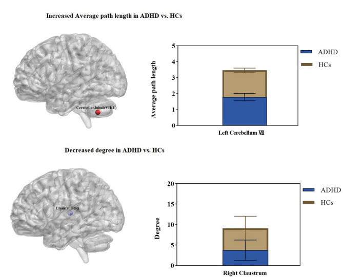
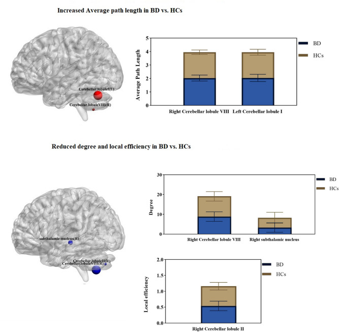
Results: Key findings show that in ADHD, increased APL in left cerebellar lobule VII indicates disrupted information flow and emotional processing, while decreased connectivity in the right claustrum may impair integration and working memory. In BD, reduced LE in right cerebellar lobule II is linked to attention and motor control deficits; increased APL in lobules I and VIII suggests disrupted network communication and emotional processing; and decreased connectivity in the right subthalamic nucleus and lobule VIII may contribute to mood swings and attention problems. In SZ, decreased LE in right putamen and left cerebellar lobule VIII relates to working memory and emotional processing deficits; reduced APL in right caudate and cerebellar lobule II implies more effort for regional communication; and increased connectivity in the caudate and right cerebellar lobules I and II likely reflects compensatory or pathological hyperactivity. Comparisons indicate SZ shows increased connectivity in the claustrum and cerebellar lobule I, unlike ADHD which shows decreases in these areas; SZ has lower network efficiency but higher caudate connectivity than BD, which has more cerebellar and subthalamic disruptions; and BD shows decreased connectivity in the claustrum and subthalamic nucleus compared to ADHD, which has more cerebellar and attention network changes.
Conclusion: These findings suggest that the PMN, particularly its segregation and integration properties, plays a key role in explaining cognitive deficits in ADHD, BD, and SZ.
Clinical trial number: Not applicable.
Supplementary Information: The online version contains supplementary material available at 10.1186/s12868-025-00979-z.
Keywords: Brain mapping; Cognitive dysfunction; Functional connectivity; Graph theory; Procedural memory; Resting-state fMRI.
Conflict of interest statement
Declarations. Ethics approval and consent to participate: The UCLA Consortium for Neuropsychiatric Phenomics (CNP) dataset [29] was utilized in this study. This dataset is publicly accessible from the OpenNeuro repository. Ethical approval for this study was obtained from the Kermanshah University of Medical Sciences Ethics Committee (reference number IR.KUMS.REC.1402.036). Consent for publication: Not applicable. Competing interests: The authors declare no competing interests.
Figures


Similar articles
-
Investigating resting-state functional connectivity changes within procedural memory network across neuropsychiatric disorders using fMRI.BMC Med Imaging. 2025 Jan 13;25(1):18. doi: 10.1186/s12880-024-01527-7. BMC Med Imaging. 2025. PMID: 39806317 Free PMC article.
-
Meso-scale network analysis of resting state-fMRI brain network connectivity performs poorly as a prognostic tool in critically ill traumatic brain injury patients.Neuroimage Rep. 2022 Jan 10;2(1):100079. doi: 10.1016/j.ynirp.2022.100079. eCollection 2022 Mar. Neuroimage Rep. 2022. PMID: 40568040 Free PMC article.
-
Spectral graph model for fMRI: A biophysical, connectivity-based generative model for the analysis of frequency-resolved resting-state fMRI.Imaging Neurosci (Camb). 2024 Dec 9;2:imag-2-00381. doi: 10.1162/imag_a_00381. eCollection 2024. Imaging Neurosci (Camb). 2024. PMID: 40800294 Free PMC article.
-
Review study of alteration functional activities and networks in ulcerative colitis using resting-state fMRI.Front Neurol. 2025 Aug 19;16:1608371. doi: 10.3389/fneur.2025.1608371. eCollection 2025. Front Neurol. 2025. PMID: 40904825 Free PMC article. Review.
-
Altered brain connectivity in hyperkinetic movement disorders: A review of resting-state fMRI.Neuroimage Clin. 2023;37:103302. doi: 10.1016/j.nicl.2022.103302. Epub 2022 Dec 24. Neuroimage Clin. 2023. PMID: 36669351 Free PMC article.
References
-
- Solmi M, Seitidis G, Mavridis D, Correll CU, Dragioti E, Guimond S, et al. Incidence, prevalence, and global burden of schizophrenia - data, with critical appraisal, from the global burden of disease (GBD) 2019. Mol Psychiatry. 2023;28:5319–27. - PubMed
-
- Kaiser ML, Schoemaker MM, Albaret JM, Geuze RH. What is the evidence of impaired motor skills and motor control among children with attention deficit hyperactivity disorder (ADHD)? Systematic review of the literature. Res Dev Disabil. 2015;36:338–57. - PubMed
Grants and funding
LinkOut - more resources
Full Text Sources
