

In-depth evaluation of XPR1 as a new prognostic indicator for endometrial cancer
- PMID: 40898098
- PMCID: PMC12403429
- DOI: 10.1186/s12885-025-14818-1
In-depth evaluation of XPR1 as a new prognostic indicator for endometrial cancer
Abstract
Background: XPR1 is crucial in the development of some tumors, yet its association with endometrial cancer (EC) remains uncertain. We propose that XPR1 exhibits elevated expression in EC and is significantly linked to unfavorable patient outcomes, positioning it as a prospective prognostic biomarker.
Methods: This study investigated XPR1 expression in 554 Uterine Corpus Endometrial Carcinoma(UCEC) cases and 35 normal tissue samples using the The Cancer Genome Atlas(TCGA) database. Western blotting confirmed XPR1 protein presence in EC cell lines (ECC-1) and normal endometrial cells (EEC). The impact of XPR1 on EC cell proliferation and invasion was assessed using EdU proliferation and Transwell invasion assays. A Dot blot assay evaluated m6A methylation of XPR1 in ECC-1 and EEC. The relationship between XPR1 and EC patient prognosis was analyzed using Kaplan-Meier survival and Cox regression analyses. A nomogram was developed to predict 1-, 3-, and 5-year survival probabilities for EC patients, with its accuracy assessed via a calibration curve. Functional enrichment analyses using Gene Ontology (GO) and Kyoto Encyclopedia of Genes and Genomes (KEGG) were conducted to elucidate biological roles and pathways related to XPR1-associated genes. The 'corrplot' R package analyzed the correlation between XPR1 expression and m6A methylation in EC, as well as its link to immune infiltration. Statistical analyses were performed using R software, with a bilateral p-value < 0.05 considered statistically significant.
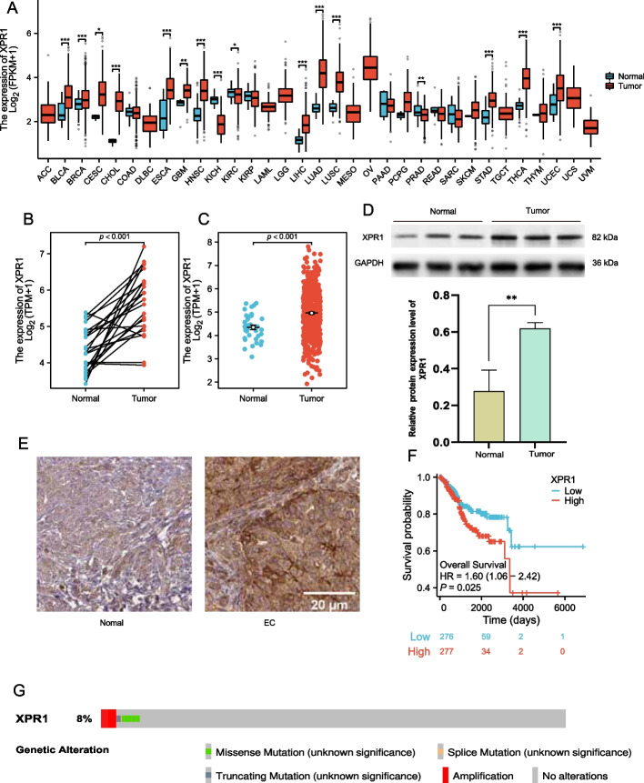
Results: This study revealed that XPR1 expression was significantly elevated in EC tissues compared to normal tissues (p < 0.001). Its expression correlated with clinical characteristics including patient age, BMI, clinical stage, histological grade, and tumor invasiveness, suggesting its potential as a prognostic marker.Kaplan-Meier analysis demonstrated that high XPR1 expression correlated with reduced overall survival (HR = 1.60, 95% CI: 1.06-2.42, p = 0.025). However, multivariable Cox regression analysis did not identify XPR1 as an independent prognostic factor.Functional experiments indicated that elevated XPR1 expression promoted proliferative and invasive capacities in endometrial carcinoma cells. XPR1 expression also associated with infiltration levels of immune cells (B cells, CD8 + T cells, CD4 + T cells, macrophages, neutrophils, and dendritic cells), suggesting potential involvement in tumor immune microenvironment regulation.Co-expressed genes with XPR1 were enriched in key biological processes including RNA processing, DNA metabolic regulation, and KEGG signaling pathways. Furthermore, XPR1 showed positive correlations with multiple m6A-related genes, and high XPR1 expression in EC coincided with elevated m6A methylation levels. Although these findings indicate that there is a correlation between XPR1 and m6A modification, it is uncertain that XPR1 can directly regulate m6A modification. The specific mechanism of XPR1 in m6A mediated post transcriptional regulation needs further study.
Conclusion: This study identified a significant association between elevated XPR1 expression and adverse clinical outcomes in endometrial carcinoma patients, with additional connections observed to m6A modification and tumor immune microenvironment dynamics. As a novel prognostic indicator, XPR1 demonstrated correlations with tumor aggressiveness features and biological processes-including cellular proliferation, invasion, and m6A/immune-related pathways-suggesting its potential biological relevance in endometrial carcinogenesis. However, its independent prognostic value remains undefined, and direct mechanistic roles in m6A-mediated post-transcriptional modulation warrant subsequent experimental validation.
Keywords: Endometrial cancer; Immune infiltration; M6A; XPR1.
© 2025. The Author(s).
Conflict of interest statement
Declarations. Ethics approval and consent to participate: Ethical approval was not necessary for this study as it did not involve human or animal subjects. Consent for publication: Not applicable. Competing interests: The authors declare no competing interests.
Figures


Similar articles
-
Expression and prognostic value of EGF-like domain multiple 6 in uterine corpus endometrial carcinoma and its correlation with immune cell infiltration.Sci Rep. 2025 Jul 2;15(1):22620. doi: 10.1038/s41598-025-07379-7. Sci Rep. 2025. PMID: 40595086 Free PMC article.
-
Comprehensive pan-cancer analysis reveals NTN1 as an immune infiltrate risk factor and its potential prognostic value in SKCM.Sci Rep. 2025 Jan 25;15(1):3223. doi: 10.1038/s41598-025-85444-x. Sci Rep. 2025. PMID: 39863609 Free PMC article.
-
Prognostic significance and immune infiltration analysis of HMGA2 in endometrial cancer.Front Immunol. 2025 Jul 9;16:1559278. doi: 10.3389/fimmu.2025.1559278. eCollection 2025. Front Immunol. 2025. PMID: 40703517 Free PMC article.
-
Cost-effectiveness of using prognostic information to select women with breast cancer for adjuvant systemic therapy.Health Technol Assess. 2006 Sep;10(34):iii-iv, ix-xi, 1-204. doi: 10.3310/hta10340. Health Technol Assess. 2006. PMID: 16959170
-
The Impact of High lncRNA Expression on Clinicopathological Characteristics and Prognosis of Endometrial Cancer Patients: A Meta-Analysis.Cancer Med. 2025 Mar;14(5):e70755. doi: 10.1002/cam4.70755. Cancer Med. 2025. PMID: 40070309 Free PMC article.
References
-
- Javadian P, Nezhat F. Endometrial Carcinoma and its Precursors. Adv Exp Med Biol. 1242:59–72. - PubMed
-
- Chen YL, Cheng WF, Lin MC, Huang CY, Hsieh CY, Chen CA. Concurrent endometrial carcinoma in patients with a curettage diagnosis of endometrial hyperplasia. J Formos Med Assoc. 2009;108(6):502–7. - PubMed
-
- Guo S, Yang J, Wu M, Xiao G. Clinical value screening, prognostic significance and key pathway identification of miR-204-5p in endometrial carcinoma: A study based on the Cancer Genome Atlas (TCGA), and bioinformatics analysis. Pathol Res Pract. 2019;215(5):1003–11. - PubMed
MeSH terms
Substances
Grants and funding
LinkOut - more resources
Full Text Sources
Research Materials
