. 2025 Aug 28;31(32):104277.
doi: 10.3748/wjg.v31.i32.104277.

Sex based relative expression of estrogen receptors and tumor necrosis factor-alpha in liver affects hepatitis C virus viral pathogenesis

Affiliations

Sex based relative expression of estrogen receptors and tumor necrosis factor-alpha in liver affects hepatitis C virus viral pathogenesis

Sarah Groover et al. World J Gastroenterol. .

Abstract

Background: Hepatocellular carcinoma (HCC) is a global health concern, representing the second most common cause of malignancy-related mortality in the world. The primary cause of HCC in the United States is chronic infection with the hepatitis C virus (HCV). Clinical observations have established sex-based differences in HCV infection with the disease progressing more severely and more rapidly in males and postmenopausal females compared to premenopausal females, suggesting that estrogens and their receptors may play an important role in hepatic defenses and development of HCV-mediated HCC. However, the precise mechanism of estrogen protection and their effects on inflammation is poorly understood.

Aim: To determine whether estrogen receptor (ER) expression is correlated with the expression of tumor necrosis factor-alpha (TNF-α) in males and females with HCV-associated diseases.

Methods: The role of ERs in modulating innate immune responses was investigated using human liver tissues with HCV/cirrhosis and HCV/HCC. Messenger RNA (mRNA) and protein (nuclear and cytoplasmic) expression were measured for all markers of interest and compared to normal human liver tissue samples.

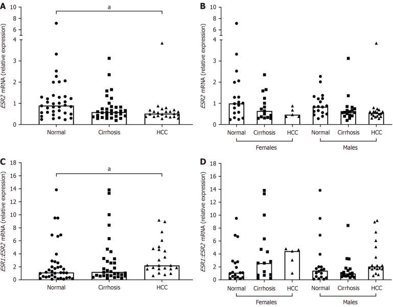
Results: ERβ was reported for the first time to have a greater mRNA expression than ERα in normal liver (P ≤ 0.001). In addition, ERβ mRNA expression was found to be decreased in diseased livers (P ≤ 0.05), while TNF-α expression was increased (P ≤ 0.0001). Upon stratifying by sex within each disease group, ESR1 was found to be negatively correlated with ESR2 in females with HCV/cirrhosis (r = -0.84, P ≤ 0.001), whereas males with HCV/cirrhosis were found to have a significant positive correlation (r = 0.57, P ≤ 0.05). ESR2 mRNA expression had a significant positive correlation with TNF-α in both HCV/cirrhosis (r = 0.61, P ≤ 0.001) and HCV/HCC patients (r = 0.45, P ≤ 0.05).

Conclusion: All together, these findings indicate that changes in ERβ and TNF-α expression are associated with worsening disease, and may be part of the sex-dependent factors in HCC pathogenesis.

Keywords: Estrogen receptor; Hepatitis C virus; Hepatocellular carcinoma; Inflammation; Liver cirrhosis; Tumor necrosis factor alpha.


Conflict of interest statement

Conflict-of-interest statement: The authors declare that they have no conflict of interest.

Figures

Figure 1
Figure 1
Message RNA expression of estrogen receptor α and estrogen receptor β in human liver tissues from normal subjects. A: Estrogen receptor subtype expression was analyzed in human liver tissues by quantitative reverse transcription polymerase chain reaction using gene specific primers. Target gene expression was normalized to GUSB and SRSF4 expression using the ΔCt method and plotted; B: Stratification of A by sex; C: Ratio of ESR1 to ESR2 message RNA expression in normals stratified by sex. Column bars represent the median. Each symbol represents one individual. Data were analyzed by Mann-Whitney U or Kruskal-Wallis test where aP ≤ 0.05. bP ≤ 0.01. dP ≤ 0.0001. mRNA: Message RNA.
Figure 2
Figure 2
Expression of estrogen receptor α in human liver tissues with hepatitis C virus-related diseases. Estrogen receptor (ER) α message RNA and protein expressions were analyzed in hepatitis C virus (HCV)-related cirrhosis and HCV-related hepatocellular carcinoma liver tissues and compared to normals. A: ESR1 transcript levels by quantitative reverse transcription polymerase chain reaction normalized to GUSB and SRSF4 expression and compared using the ΔΔCt method; B: Stratification of A by sex; C: ERα protein expression by Western blot using whole liver tissue lysates (tot); D: Quantification of C by densitometry from multiple blots and normalized to total protein; E: ERα protein expression in human liver tissues stratified by sex. Column bars represent the median. Each symbol represents one individual. Data were analyzed by Kruskal-Wallis test with Dunn’s Multiple Comparisons posttest where P ≤ 0.05 was considered significant. mRNA: Message RNA; HCC: Hepatocellular carcinoma; ER: Estrogen receptor; TPS: Total protein stain.
Figure 3
Figure 3
Message RNA expression of estrogen receptor β in human liver tissues with hepatitis C virus-related diseases. Estrogen receptor (ER) β message RNA (mRNA) expression was analyzed in hepatitis C virus (HCV)-related cirrhosis and HCV-related hepatocellular carcinoma liver tissues and compared to normals. A: ESR2 transcript levels by quantitative reverse transcription polymerase chain reaction normalized to GUSB and SRSF4 expression and compared using the ΔΔCt method; B: Stratification of A by sex; C: Ratio of ESR1 to ESR2 mRNA expression; D: Stratification of C by sex. Column bars represent the median. Each symbol represents one individual. Data were analyzed by Kruskal-Wallis test with Dunn’s Multiple Comparisons posttest where P ≤ 0.05 was considered significant. aP ≤ 0.05. mRNA: Message RNA; HCC: Hepatocellular carcinoma.
Figure 4
Figure 4
Protein expression of estrogen receptor β in human liver tissues with hepatitis C virus-related diseases. Estrogen receptor (ER) β protein expression was analyzed in hepatitis C virus (HCV)-related cirrhosis and HCV-related hepatocellular carcinoma liver tissues and compared to normals. A: ERβ protein expression by Western blot in human liver tissues fractionated by nuclear and cytoplasmic protein; B and C: Quantification of A by densitometry from multiple blots and normalized to total protein; D and E: ERβ protein expression in nuclear and cytoplasmic fractions of human liver tissues and stratified by sex. Column bars represent the median. Each symbol represents one individual. Data were analyzed by Kruskal-Wallis test with Dunn’s Multiple Comparisons posttest where aP ≤ 0.05. bP ≤ 0.01. cP ≤ 0.001. Nuc: Nuclear; Cyt: Cytoplasmic; HCC: Hepatocellular carcinoma; ER: Estrogen receptor; TPS: Total protein stain.
Figure 5
Figure 5
Expression of tumor necrosis factor-alpha in human liver tissues with hepatitis C virus-related diseases. Tumor necrosis factor-alpha (TNF-α) message RNA and protein expressions were analyzed in hepatitis C virus (HCV)-related cirrhosis and HCV-related hepatocellular carcinoma liver tissues and compared to normals. A: TNF-α transcript levels by quantitative reverse transcription polymerase chain reaction normalized to GUSB and SRSF4 expression and compared using the ΔΔCt method; B: Stratification of A by sex; C: TNF-α protein expression by Western blot using whole liver tissue lysates; D: Quantification of C by densitometry from multiple blots and normalized to total protein; E: Stratification of D by sex. Column bars represent the median. Each symbol represents one individual. Data were analyzed by Kruskal-Wallis test with Dunn’s Multiple Comparisons posttest where aP ≤ 0.05. cP ≤ 0.001. TNF-α: Tumor necrosis factor-alpha; HCC: Hepatocellular carcinoma; TPS: Total protein stain.

References

    1. Mittal S, El-Serag HB. Epidemiology of hepatocellular carcinoma: consider the population. J Clin Gastroenterol. 2013;47 Suppl:S2–S6. - PMC - PubMed
    1. United States Cancer Statistics. About the U.S. Cancer Statistics Data Visualizations Tool. [cited July 1, 2025]. Available from: https://www.cdc.gov/united-states-cancer-statistics/dataviz/index.html .
    1. Hofmeister MG, Rosenthal EM, Barker LK, Rosenberg ES, Barranco MA, Hall EW, Edlin BR, Mermin J, Ward JW, Ryerson AB. Estimating Prevalence of Hepatitis C Virus Infection in the United States, 2013-2016. Hepatology. 2019;69:1020–1031. - PMC - PubMed
    1. Dubuisson J, Cosset FL. Virology and cell biology of the hepatitis C virus life cycle: an update. J Hepatol. 2014;61:S3–S13. - PubMed
    1. Hoofnagle JH, Mullen KD, Jones DB, Rustgi V, Di Bisceglie A, Peters M, Waggoner JG, Park Y, Jones EA. Treatment of chronic non-A,non-B hepatitis with recombinant human alpha interferon. A preliminary report. N Engl J Med. 1986;315:1575–1578. - PubMed

MeSH terms