. 2025 Aug 18:15:1617101.
doi: 10.3389/fcimb.2025.1617101. eCollection 2025.

EPEC autotransporter adhesin (Eaa): a novel adhesin identified in atypical enteropathogenic Escherichia coli

Affiliations

EPEC autotransporter adhesin (Eaa): a novel adhesin identified in atypical enteropathogenic Escherichia coli

Henrique Orsi et al. Front Cell Infect Microbiol. .

Abstract

Enteropathogenic Escherichia coli (EPEC) is a pathogen that causes diarrhea that can be subdivided into typical (tEPEC) and atypical (aEPEC), based on the production of an adhesin termed Bundle-Forming Pilus (BFP) in the former group. aEPEC is one of the main bacterial pathogens isolated from individuals with diarrhea, and some serotypes have been implicated in diarrheal outbreaks in Brazil, such as the O2:H16. A comparative genomic analysis of aEPEC of this serotype led to the identification of a gene encoding a previously uncharacterized autotransporter protein. In the present study, this novel autotransporter protein was characterized and named EPEC Autotransporter Adhesin (Eaa). The Eaa-encoding gene (eaa) is located in a chromosomal prophage region of 17,014 base pairs, organized in 20 open reading frames and inserted downstream to the threonine-tRNA. A recombinant plasmid termed pIC (pBAD/Myc-His A harboring the eaa gene from aEPEC BA92) was transformed in the MS427 host bacteria, and the MS427(pIC) was used in phenotypic assays. Immunogold-labelling transmission electron microscopy, using anti-Eaa antibodies, showed the presence of Eaa in the cell surface of the wild-type BA92 and MS427(pIC) strains. Subsequently, we demonstrated that Eaa mediates bacterial autoaggregation, biofilm formation and binding to several components of the extracellular matrix, including fibrinogen, plasma and cellular fibronectin, type I, III as well as V collagen and laminin. In summary, we demonstrated that Eaa harbors several adherence properties and may contribute to the pathogenicity of some aEPEC isolates by mediating the interaction of this pathogen with biotic and abiotic surfaces.

Keywords: adhesin; atypical EPEC; autotransporter protein; bacterial pathogenesis; biofilm; fibronectin.


Conflict of interest statement

The authors declare that the research was conducted in the absence of any commercial or financial relationships that could be construed as a potential conflict of interest.

Figures

Figure 1
Figure 1
Visualization of the chromosomal region that harbors the eaa gene in the aEPEC BA92 strain. A comparative genomic analysis of the chromosome of the aEPEC BA92 (NCBI accession number: CP176406) with the draft genome sequence of the aEPEC IAL5132 (NCBI accession number: PIKD01000024.1, contig No. 24) revealed that the eaa gene is located on a chromosomal region with 17,014 base pairs organized in 20 coding sequences. This chromosomal region is inserted downstream to the gene encoding the transfer RNA for the amino acid threonine (tRNA-Thr). The protein ID for the Eaa is XWX38401.1.
Figure 2
Figure 2
Protein domains and tertiary structure of the Eaa protein. This image illustrates known protein domains found in Eaa according to InterPro and Pfam databases. (A) and structural prediction according to ColabFold (B, C). (A) the signal peptide, four AIDA domain repeats, a pertactin domain and the translocator domain (β- barrel) are highlighted. (B), the N- and C-terminal regions are respectively shown in blue and red, while in (C) the protein is colored according to its lDDT (local Distance Difference Test) score which is categorized as follows: Dark blue >90, light blue = 80, green = 70, yellow = 60 and red <50.
Figure 3
Figure 3
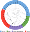
The novel autotransporter protein Eaa belongs to the AIDA-I family of adhesins. The amino acid sequences of several well-characterized autotransporter proteins ( Supplementary Table S1 ), were aligned using ClustalW in MEGA 11 and the maximum likelihood tree was calculated using IQ-Tree 1.6.12 along with the UFBoot2 algorithm using 1,000 bootstraps and the PMB model. The resulting Newick file was visualized using the iTOL software. The maximum likelihood tree demonstrated that Eaa belongs to the AIDA-I family and is closely related to the adhesins TibA and EhaD, first described in the enterotoxigenic (ETEC) and enterohemorrhagic (EHEC) E. coli pathotypes, respectively. The nodes with ultrafast bootstrap support values >80% are labeled with a black circle. The novel autotransporter protein Eaa is highlighted in bold.
Figure 4
Figure 4
Identification of Eaa on the bacterial cell surface of aEPEC BA92 and MS427(pIC) strains. Immunogold-labelling (IGL) preparations using pre-immune (A-C) or anti-Eaa (D-F) sera, produced in rabbit (primary antibodies), and goat anti-rabbit serum conjugated with 10 nm colloidal gold particles (secondary antibody) were analyzed by transmission electron microscopy. IGL using anti-Eaa serum (D-F), demonstrated that Eaa is present on the bacterial cell surface of both BA92 (wild- type) and MS427(pIC) strains, as indicated by arrows (D, F). Note that the preparations were not negatively stained, and that gold particles were not observed on the surface of the MS427(pBAD) strain (E), used as a negative control in this assay, nor in any of the preparations incubated with the pre-immune serum (A-C). Bars, 200 nm.
Figure 5
Figure 5
Eaa promotes bacterial autoaggregation. The autoaggregation assay was carried out under two experimental conditions: (A) with homogenization of the bacterial cultures and (B) without homogenization of the bacterial cultures before reading the optical density at 600 nm (OD600). Note, in panel (B), that the OD600 of the bacteria that produce Eaa, MS427(pIC), as well as the Antigen 43, MS427(pCO4), progressively decrease throughout the autoaggregation assay. Statistical difference between MS427(pBAD), used as a negative control in this assay, and MS427(pIC) was observed at all time points (P<0.001). Error bars show standard deviation.
Figure 6
Figure 6
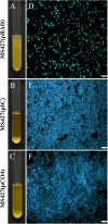
Eaa promotes the formation of large bacterial aggregates. (A-C) show the settling patterns of the cultures left statically for 2 hours at room temperature, while the (D-F) are fluorescence microscope images of bacterial sediments stained with DAPI. Note in panels (B, C) the large amount of bacterial cells sedimented at the bottom of the test tube from the broth cultures of the strains MS472(pIC) and MS427(pCO4), that produce the autotransporter proteins Eaa and Antigen 43, respectively. The bacterial aggregates induced by the production of Eaa and Antigen 43 can be visualized in (E, F), respectively, while in (D) only the occurrence of isolated bacteria is observed. Bars = 10 µm.
Figure 7
Figure 7
Eaa increases biofilm formation on polystyrene. Biofilm production was evaluated on polystyrene surface in three periods of time: 24, 48 and 72 hours. Note that the bacteria producing the autotransporter proteins Eaa, MS427(pIC), as well as the Antigen 43, MS427(pCO4), produce significantly more biofilm than the host bacteria carrying the pBAD/Myc-His A expression vector. Error bars show standard deviation. ****P<0.0001 and ns, non-significant.
Figure 8
Figure 8
Recombinant Eaa binds to several plasma and extracellular matrix (ECM) components. For this assay, wells were coated with 1 μg of the ECM components followed by the addition of 0.1 μM of recombinant Eaa per well. The binding assay to ECM components demonstrated that the recombinant Eaa-His protein binds to fibrinogen, plasma and cellular fibronectin, type I, III and V collagen and laminin. Bovine serum albumin (BSA, nonglycosylated attachment-negative control protein) and fetuin (highly glycosylated attachment-negative control protein) were used as negative controls in this binding assay. Optical densities were taken at 492 nm. Data represent the mean ± standard error of three independent experiments. Statistical differences relative to the values obtained for BSA are shown. **P=0.0097, ***P=0.0003 and ****P<0.0001.
Figure 9
Figure 9
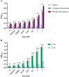
Eaa dose-dependently binds to cellular and plasma fibronectin through its heparin-binding domain (F30). Binding of recombinant Eaa (0 to 2 µM) to cellular and plasma fibronectin (A), as well as to F30 (Heparin Binding Domain, HBD) and F45 (Gelatin Binding Domain, GBD) fibronectin domains (B) was assessed by an ELISA- based assay. Wells were coated with 1 µg of intact fibronectin (A) or with F30 and F45 fragments (B) and increasing amounts of recombinant Eaa (0 – 2 µM) were added to each well. Optical densities were taken at 492 nm. Note that the recombinant Eaa-His protein binds to cellular as well as plasma fibronectin in a dose-dependent manner (A); and binds more efficiently to the F30 fibronectin domain (B). Data represents the mean ± standard error of three independent experiments. Statistical differences relative to the values obtained for fetuin are shown in (A). Statistical comparisons were performed using multiple t tests. **P ≤ 0.01; ***P ≤ 0.001; ****P ≤ 0.0001 and ns, non-significant.

References

    1. Abe C. M., Salvador F. A., Falsetti I. N., Vieira M. A. M., Blanco J., Blanco J. E., et al. (2008). Uropathogenic Escherichia coli (UPEC) strains may carry virulence properties of diarrhoeagenic E. coli. FEMS Immunol. Med. Microbiol. 52, 397–406. doi: 10.1111/j.1574-695X.2008.00388.x, PMID: - DOI - PubMed
    1. Abe C. M., Trabulsi L. R., Blanco J., Blanco M., Dhabi G., Blanco J. E., et al. (2009). Virulence features of atypical enteropathogenic Escherichia coli identified by the eae(+) EAF-negative stx(-) genetic profile. Diagn. Microbiol. Infect. Dis. 64, 357–365. doi: 10.1016/j.diagmicrobio.2009.03.025, PMID: - DOI - PubMed
    1. Abreu A. G., Bueris V., Porangaba T. M., Sircili M. P., Navarro-Garcia F., Elias W. P. (2013). Autotransporter protein-encoding genes of diarrheagenic Escherichia coli are found in both typical and atypical enteropathogenic E. coli strains. Appl. Environ. Microbiol. 79, 411–414. doi: 10.1128/AEM.02635-12, PMID: - DOI - PMC - PubMed
    1. Afset J. E., Bevanger L., Romundstad P., Bergh K. (2004). Association of atypical enteropathogenic Escherichia coli (EPEC) with prolonged diarrhoea. J. Med. Microbiol. 53, 1137–1144. doi: 10.1099/jmm.0.45719-0, PMID: - DOI - PubMed
    1. Allsopp L. P., Beloin C., Ulett G. C., Valle J., Totsika M., Sherlock O., et al. (2012). Molecular characterization of UpaB and UpaC, two new autotransporter proteins of uropathogenic Escherichia coli CFT073. Infect. Immun. 80, 321–332. doi: 10.1128/IAI.05322-11, PMID: - DOI - PMC - PubMed

MeSH terms

LinkOut - more resources