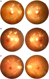
Case Report: Concurrent retinal vasculitis and optic neuritis in systemic lupus erythematosus
- PMID: 40901471
- PMCID: PMC12399518
- DOI: 10.3389/fimmu.2025.1646850
Case Report: Concurrent retinal vasculitis and optic neuritis in systemic lupus erythematosus
Abstract
Systemic lupus erythematosus (SLE) is a multisystem autoimmune disease that can affect the ocular system, with retinal vasculitis and optic neuritis being rare but serious manifestations. We present a case of a 26-year-old female with newly diagnosed SLE who developed both retinal vasculitis and optic neuritis, leading to progressive visual impairment. She was successfully treated with methylprednisolone and rituximab, achieving significant visual recovery. A review of existing literature highlights the diagnostic challenges, pathophysiology, and optimal treatment strategies for such cases. Our findings emphasize the importance of early recognition and aggressive immunosuppressive therapy in improving patient outcomes.
Keywords: autoimmune ocular disease; immunosuppressive therapy; optic neuritis; retinal vasculitis; systemic lupus erythematosus.
Copyright © 2025 Jin, Liu, Wang, Fang, Nie, Li, Li and Li.
Conflict of interest statement
The authors declare that the research was conducted in the absence of any commercial or financial relationships that could be construed as a potential conflict of interest.
Figures


Similar articles
-
Optic neuritis and retinal vasculitis as primary manifestations of systemic lupus erythematosus.Med J Malaysia. 2002 Dec;57(4):490-2. Med J Malaysia. 2002. PMID: 12733176
-
Neuro-ophthalmologic manifestations of systemic lupus erythematosus: a systematic review.Int J Rheum Dis. 2014 Jun;17(5):494-501. doi: 10.1111/1756-185X.12337. Epub 2014 Mar 28. Int J Rheum Dis. 2014. PMID: 24673755
-
Cyclophosphamide versus methylprednisolone for treating neuropsychiatric involvement in systemic lupus erythematosus.Cochrane Database Syst Rev. 2013 Feb 28;2013(2):CD002265. doi: 10.1002/14651858.CD002265.pub3. Cochrane Database Syst Rev. 2013. PMID: 23450535 Free PMC article.
-
Cyclophosphamide versus methylprednisolone for treating neuropsychiatric involvement in systemic lupus erythematosus.Cochrane Database Syst Rev. 2006 Apr 19;(2):CD002265. doi: 10.1002/14651858.CD002265.pub2. Cochrane Database Syst Rev. 2006. Update in: Cochrane Database Syst Rev. 2013 Feb 28;(2):CD002265. doi: 10.1002/14651858.CD002265.pub3. PMID: 16625558 Updated.
-
Treatment of systemic lupus erythemus overlap syndrome with autoimmune hepatitis using a combination of glucocorticoids and immunosuppressive agents: Case report.Medicine (Baltimore). 2025 Aug 1;104(31):e43570. doi: 10.1097/MD.0000000000043570. Medicine (Baltimore). 2025. PMID: 40760563 Free PMC article.
References
Publication types
MeSH terms
Substances
LinkOut - more resources
Full Text Sources
Medical

