Review
. 2025 Aug 27;17(8):107041.
doi: 10.4254/wjh.v17.i8.107041.

Imaging insights into pediatric liver masses: A comprehensive minireview for hepatology practice

Affiliations
Review

Imaging insights into pediatric liver masses: A comprehensive minireview for hepatology practice

Mina'a Shahid et al. World J Hepatol. .

Abstract

Pediatric liver masses encompass a diverse spectrum of benign and malignant lesions, with distinct patterns based on patient age. Optimal imaging is critical for timely diagnosis, management, and prognosis. This pictorial minireview categorizes pediatric liver masses by age group to guide hepatology and radiology practice, with an emphasis on imaging characteristics. In children from birth to six years of age, the most common liver masses include hepatoblastoma, the most common primary hepatic malignancy in this age group; infantile hemangioma, a benign vascular tumor with a characteristic appearance on imaging; and mesenchymal hamartoma, a rare developmental lesion. For children older than six years, liver masses are distinct, with hepatocellular carcinoma being the predominant malignant lesion. Benign masses such as focal nodular hyperplasia and hepatocellular adenoma also emerge in this age range, often linked to hormonal influences or metabolic disorders. The masses observed across all pediatric age groups include hepatic cysts, choledochal cysts, hydatid cysts, pyogenic and amebic abscesses, tuberculosis, lymphoma, and metastases, each presenting with unique imaging features essential for differential diagnosis. This minireview provides a comprehensive, age-based overview of pediatric liver masses, focusing on clinical presentation and key imaging findings to support accurate diagnosis and optimize management strategies in clinical hepatology, particularly in low resource settings.

Keywords: Benign liver lesions; Hepatic masses; Malignant liver lesions; Pediatric hepatic imaging; Pediatric liver tumors.


Conflict of interest statement

Conflict-of-interest statement: The authors declare no conflict of interest related to this article.

Figures

Figure 1
Figure 1
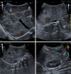
Hepatoblastoma in a four-year-old child with a tense abdomen, weight loss and lethargy. A: Color Doppler examination; B: Right lobe of the liver. Ultrasound shows a large, solid, heterogeneous, predominantly hyperechoic lesion in the right lobe of the liver with internal vascularity on color Doppler examination (arrows).
Figure 2
Figure 2
Hepatoblastoma in a six-month-old boy who presented with complaints of abdominal distension for two weeks. A: Axial computed tomography (CT) image; B: Coronal CT image; C: Sagittal CT image. They show an enlarged liver with a large heterogeneous lesion occupying both lobes of the liver (arrow in A), involving segments I, IV, V and VIII. It displaces the left portal vein (arrow in B) and has internal low attenuation areas representing necrosis (asterisk in image C). No internal calcifications are noted. This was staged as pretreatment extent of disease III C (caudate).
Figure 3
Figure 3
Hepatoblastoma in a two-year-old boy with an abdominal mass and increased alpha-fetoprotein levels. A and B: Axial T2-weighted images from an magnetic resonance imaging abdomen show heterogeneous lesions (arrows) involving the right lobe of the liver, predominantly segments VI and V; C and D: Axial T1 post-contrast images. The lesions show heterogeneous postcontrast enhancement (arrows). There is no infiltration of the mass into the inferior vena cava, or portal or hepatic veins. This was staged as pretreatment extent of disease II F (multifocal) disease involving right anterior and posterior sections.
Figure 4
Figure 4
Congenital hepatic hemangioma in an eleven-month-old boy who presented with complaints of vomiting and a mass in the right hypochondrium. A: Ultrasound shows a large heterogeneous lesion in the right lobe with internal calcifications and hypoechoic areas (arrows); B and C: Vascularity is noted within the lesion on Doppler with both arterial and venous flow on spectral Doppler, with low arterial resistive indices.
Figure 5
Figure 5
Congenital hepatic hemangioma in an eleven-month-old boy who presented with complaints of vomiting and a mass in the right hypochondrium. A-C: Axial computed tomography (CT) images; D-F: Coronal CT images. They show a large heterogeneous lesion in the right lobe of liver, with peripheral nodular discontinuous arterial enhancement and gradual central filling on venous and delayed phases (arrows in A-C).
Figure 6
Figure 6
Congenital hepatic hemangioma in a six- month-old boy who presented with complaints of a right hypochondrium mass. A: Axial T2-weighted image; B: Coronal T2-weighted image; C: T1-weighted image; D: T1 postcontrast image. Images from an magnetic resonance imaging abdomen show a large solid hyperintense lesion on T2 (arrows) involving segments V, VII and VIII of the liver, which appears hypointense on T1-weighted images and shows avid enhancement on postcontrast images.
Figure 7
Figure 7
A 6-month-old girl with multifocal infantile hepatic hemangioma who was managed with propranolol. A: Grayscale ultrasound (US) image; B: Color Doppler US image. Grayscale and color Doppler US images taken 5 months apart show multiple hypoechoic lesions in both lobes of the liver (arrows in A), with marked interval regression in B.
Figure 8
Figure 8
Mesenchymal hamartoma of the liver in a 1-day-old baby boy with a history of a multicystic echogenic structure in the right side of the abdomen on antenatal scan. A: Grayscale ultrasound (US) image; B: Color Doppler US image. Images show a large lobulated heterogeneous multiseptated cystic lesion in the liver interspersed with solid component (arrows in A) with some internal vascularity in B.
Figure 9
Figure 9
Hepatocellular carcinoma in a 14-year-old boy with a prior history of hepatitis C virus infection and liver cirrhosis. A-C: Axial images; D: Coronal image. Images from computed tomography scan showing early arterial enhancement (arrow in A) in a lesion seen in segment VIII of the liver with washout on portal venous and delayed phases (asterisk in B and C). A large peripancreatic lymph nodal conglomerate mass is also noted in image D (curved arrow).
Figure 10
Figure 10
Fibrolamellar hepatocellular carcinoma in a 12-year-old boy who presented with an abdominal mass and jaundice. A-C: Axial computed tomography (CT) images; D and E: Coronal CT images; F: Sagittal CT image. Images show a lobulated arterially enhancing lesion (arrow in A) in the left lobe of the liver involving segment III with a hypodense central scar (arrows in D and F). The scar shows washout on venous and delayed phases (arrows in B and C). Attenuated left portal vein is seen (asterisk in E) suggesting a tumor thrombus.
Figure 11
Figure 11
Focal nodular hyperplasia in a 9-year-old boy with complaints of progressively worsening abdominal pain for one year and loose stools for five months. A and B: Axial images; C: Sagittal image; D: Coronal image. Images from computed tomography scan show a large, well-defined, exophytic, intensely enhancing hepatic lesion (asterisk in C and D) arising from the left hepatic lobe, involving segments II and III, with a central hypodense scar (arrows in A, B and D).
Figure 12
Figure 12
Hepatic adenoma in a 10-year-old boy with short stature and an enlarged liver. A: Color Doppler ultrasound (US) image; B: Grayscale US image. Images show a hyperechoic lesion (arrow) in the right lobe of the liver involving segment VI, with no discernible internal vascularity on color Doppler imaging.
Figure 13
Figure 13
Simple hepatic cyst in a 7-year-old girl who underwent grayscale ultrasound for nonspecific abdominal pain. An anechoic structure (arrow) is seen in the right lobe of the liver involving segment VI, without any internal debris or septae, which is consistent with a simple hepatic cyst.
Figure 14
Figure 14
Simple hepatic cyst in a 16-year-old girl who presented with complaints of abdominal pain. A and B: Axial computed tomography (CT) and T2-weighted magnetic resonance (MR) images; C and D: Coronal CT and T2-weighted MR images. CT images with corresponding T2-weighted images from magnetic resonance imaging show small hypodense and T2 hyperintense lesions in the right lobe of the liver, respectively, involving segment VII, which has thin walls without internal debris or septations, and no apparent biliary communication, indicating simple hepatic cysts (arrows).
Figure 15
Figure 15
Choledochal cyst in an 11-year-old girl who presented with complaints of abdominal pain and mild jaundice. A: Axial section; B: Coronal section; C: Sagittal section. Sections from the computed tomography scan show dilatation of the extrahepatic biliary duct (arrows) as well as left lobe intrahepatic channels (asterisk in B), representing a type IV choledochal cyst according to Todani classification.
Figure 16
Figure 16
Choledochal cyst in a 15-year-old girl who presented with complaints of pain in the abdomen. A two-dimensional magnetic resonance cholangiopancreatography image showing cystic and fusiform dilatation of the extrahepatic bile duct (orange arrow) and left main hepatic duct (arrowhead), representing a type IVa choledochal cyst according to Todani classification. A distended gallbladder is also noted (asterisk).
Figure 17
Figure 17
Hydatid cyst in a 15-year-old girl who presented with abdominal pain. Grayscale ultrasound images show an anechoic structure with internal floating membranes (arrows) in the right lobe of the liver, consistent with a hydatid cyst.
Figure 18
Figure 18
Liver hydatid cysts in two pediatric patients. A and B: Computed tomography (CT) scan of a 14-year-old girl who presented with abdominal pain. Axial and coronal images show a cystic lesion in the left lobe of the liver with daughter cysts and internal septa (arrows) with a spoke-wheel appearance; C and D: CT scan of an 11-year-old boy who presented with complaints of abdominal pain for one month. Axial and coronal images show a hydatid cyst involving the right lobe of the liver with detached endocysts (arrows) floating in the cyst fluid, showing the characteristic water lily sign.
Figure 19
Figure 19
A 15-year-old girl presented with hydatid cysts in the right lobe of the liver diagnosed on ultrasound. A: Aspiration; B: Injection; C: Reaspiration; D: Postprocedure film. Fluoroscopic images from percutaneous puncture, aspiration, injection and reaspiration performed in the management of the hydatid cyst with postprocedure film. The cavity was rinsed with 100 mL of 20% hypertonic saline, multiple times and 20 mL of absolute alcohol was also injected into the cyst after aspiration.
Figure 20
Figure 20
Multifocal hepatic abscesses in a 3-year-old boy who presented with complaints of abdominal pain and fever. A and B: Axial computed tomography (CT) images; C: Coronal CT image; D: Sagittal CT image. Images show a multiseptated thick-walled heterogeneous lesion demonstrating cluster sign (arrows in A-C) within segment VI of the liver with subcapsular extension (asterisks in B and D).
Figure 21
Figure 21
Pyogenic hepatic abscess in a 10-year-old girl who presented with complaints of right costal margin tenderness, fever, and vomiting. A: Axial computed tomography (CT) image; B: Coronal CT image. Images show a lobulated hypodense lesion in the right hepatic lobe segment VII with peripheral enhancement and surrounding reactive changes/hyperemia (arrows).
Figure 22
Figure 22
A 17-year-old boy with multifocal liver and bony tuberculous lesions (pathologically proven), initially thought to be metastatic disease. A and B: Axial T2-weighted magnetic resonance images showing nodular T2 hyperintense lesions in both lobes of the liver (arrows); C and D: No uptake was noted on the axial hepatocyte-specific phase (C), and minimal enhancement was seen on postcontrast T1 sequences (D); E: Multiple enhancing vertebral marrow lesions are observed on the coronal T1 postcontrast image (arrows).
Figure 23
Figure 23
Lymphomatous liver deposits in a 16-year-old boy with Hodgkin’s lymphoma (Lugano stage IV). A and B: Axial and coronal computed tomography (CT) images show multiple hypodense lesions in the liver (arrows) and spleen (arrows); C and D: The corresponding axial and coronal positron emission tomography/CT fusion images show hypermetabolic activity at the site of lesions in the liver (arrows) and spleen (arrows).
Figure 24
Figure 24
A 14-year-old girl, who was a known case of left ovarian mixed germ cell tumor (95% dysgerminoma), presented with widespread liver metastasis and hemoperitoneum. A: Axial computed tomography (CT) image; B: Coronal CT image. Images show numerous large liver lesions (arrows in A) with high-density ascites suggesting tumor rupture (arrow in B).

Similar articles

References

    1. Chiorean L, Cui XW, Tannapfel A, Franke D, Stenzel M, Kosiak W, Schreiber-Dietrich D, Jüngert J, Chang JM, Dietrich CF. Benign liver tumors in pediatric patients - Review with emphasis on imaging features. World J Gastroenterol. 2015;21:8541–8561. - PMC - PubMed
    1. El Asmar A, El Rassi Z. Hepatoblastoma in childhood, long term survival achieved: 2 case reports and literature review. Int J Surg Case Rep. 2016;21:55–58. - PMC - PubMed
    1. Lucas B, Ravishankar S, Pateva I. Pediatric Primary Hepatic Tumors: Diagnostic Considerations. Diagnostics (Basel) 2021;11:333. - PMC - PubMed
    1. Yang T, Whitlock RS, Vasudevan SA. Surgical Management of Hepatoblastoma and Recent Advances. Cancers (Basel) 2019;11:1944. - PMC - PubMed
    1. Towbin AJ, Meyers RL, Woodley H, Miyazaki O, Weldon CB, Morland B, Hiyama E, Czauderna P, Roebuck DJ, Tiao GM. 2017 PRETEXT: radiologic staging system for primary hepatic malignancies of childhood revised for the Paediatric Hepatic International Tumour Trial (PHITT) Pediatr Radiol. 2018;48:536–554. - PubMed

LinkOut - more resources