. 2025 Sep 5;11(36):eadx7172.
doi: 10.1126/sciadv.adx7172. Epub 2025 Sep 3.

Deltex and RING-UIM E3 ligases cooperate to create a ubiquitin-ADP-ribose hybrid mark on tankyrase, promoting its stabilization

Affiliations

Deltex and RING-UIM E3 ligases cooperate to create a ubiquitin-ADP-ribose hybrid mark on tankyrase, promoting its stabilization

Jerome Perrard et al. Sci Adv. .

Abstract

ADP-ribosylation can occur as mono-ADP-ribose (MAR) or be extended into poly-ADP-ribose (PAR). Tankyrase, a PAR transferase, adds PAR to itself and other proteins targeting them for proteasomal degradation via the PAR-binding E3 ligase RNF146. This degradation can be counteracted by RING-UIM E3 ligases RNF114 and RNF166, although the process is unclear. Here, we identify a mechanism that can regulate the balance between MAR and PAR on tankyrase to control degradation. We show that Deltex E3 ligases DTX2 and DTX3 catalyze monoubiquitylation of tankyrase in cells. This ubiquitylation occurs, not on a (canonical) lysine, but rather on MAR, creating a monoubiquitin-MAR hybrid mark. RNF114 and RNF166 recognize this mark using a unique hybrid reader domain and further diubiquitylate it. This ubiquitylation of MAR, which occurs near the ADP-ribose addition site, prevents PAR formation, antagonizing the action of the PAR-binding E3 ligase RNF146 and stabilizing tankyrase. These findings reveal an interplay between ubiquitin, ADP-ribose, and E3 ligases in cellular signaling.


Figures

Fig. 1.
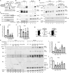
Fig. 1.. TNKS is ubiquitylated on MAR.
(A) Schematic diagram of RNF166 and the Di19-UIM construct. The pathway for RNF166-mediated K11-linked diubiquitylation of TNKS is shown below. (B) Immunoblot analysis of TNKS1/2 DKO HEK293T cells transfected with MycSAMPARP, FlagDi19-UIM, and HA-Ub plasmids; immunoprecipitated with anti-HA antibody; stained with amido black; and probed with antibodies indicated on the left. Right panel shows HA IP reprobed with anti-HA antibody. (C) Schematic diagram showing the cleavage sites for ARH3 and NH2OH on Ub-MAR-SAMPARP. Immunoblot analysis of Ub-MAR-MycSAMPARP that was HA immuno-isolated from TNKS1/2 DKO HEK293T cells transfected with MycSAMPARP, FlagD19UIM, and HA-Ub plasmids, and then incubated in vitro for 30 min at 37°C with the indicated treatments, stained with amido black, and probed with anti-Myc antibody. (D) Schematic diagram showing the cleavage sites for ARH3, NH2OH, and Cezanne on diUb-MAR-SAMPARP. Immunoblot analysis of diUb-MAR-MycSAMPARP that was HA immuno-isolated from TNKS1/2 DKO HEK293T cells transfected with MycSAMPARP, FlagRNF166, and HA-Ub plasmids and then incubated in vitro for 30 min at 37°C with the indicated treatments, stained with amido black, and probed with anti-Myc antibody. (E) Schematic diagram showing the cleavage sites for ARH3 and PARG on Ub-PAR-MycSAMPARP. Immunoblot analysis of Ub-MAR-MycSAMPARP that was HA immuno-isolated from denatured TNKS1/2 DKO HEK293T cells transfected with the MycSAMPARP, FlagD19UIM, and HA-Ub plasmids and then incubated in vitro for 30 min at 37°C with the indicated treatments; stained with amido black; and probed with antibodies indicated on the left. (F) Immunoblot analysis of TNKS1/2 DKO HEK293T cells transfected with MycSAMPARP, HA-Ub, and the indicated Flag plasmids; immunoprecipitated with anti-HA antibody; stained with amido black; and probed with the antibodies indicated on the left. (G) Schematic diagram showing K11-linked diubiquitin on MAR on TNKS. IgG, immunoglobulin G.
Fig. 2.
Fig. 2.. DTX2 monoubiquitylates TNKS on MAR.
(A) Schematic of the Deltex E3 ligase family. (B) Immunoblot analysis of TNKS1/2 DKO HEK293T cells transfected with the indicated plasmids, immunoprecipitated from denatured cell extracts with anti-HA antibody, stained with amido black, and probed with antibodies indicated on the left. (C) Immunoblot analysis of TNKS1/2 DKO HEK293T cells transfected with GFP or DTX2 siRNA and probed with antibodies indicated on the left. (D) Immunoblot analysis of TNKS1/2 DKO HEK293T cells transfected with MycSAMPARP, HA-Ub, and GFP or DTX2 siRNA; immunoprecipitated with anti-HA antibody; stained with amido black; and probed with antibodies indicated on the left. (E) Schematic of DTX2 mutant constructs. (F) Immunoblot analysis of TNKS1/2 DKO HEK293T cells transfected with the indicated plasmids, immunoprecipitated with anti-Flag antibody, stained with amido black, and probed with antibodies indicated on the left. (G) Immunoblot analysis of TNKS1/2 DKO HEK293T cells transfected with the indicated plasmids, immunoprecipitated with anti-HA antibody, stained with amido black, and probed with antibodies indicated on the left. (H) Immunoblot analysis of TNKS1/2 DKO HEK293T cells transfected with the indicated plasmids, immunoprecipitated with anti-Flag antibody, stained with amido black, and probed with antibodies indicated on the left. (I) Immunoblot analysis of TNKS1/2 DKO HEK293T cells transfected with the indicated plasmids, immunoprecipitated with anti-Flag antibody, stained with amido black, and probed with antibodies indicated on the left. (J) Immunoblot analysis of TNKS1/2 DKO HEK293T cells transfected with the indicated plasmids, immunoprecipitated with anti-HA antibody, stained with amido black, and probed with antibodies indicated on the left. (K) Immunoblot analysis of TNKS1/2 DKO HEK293T cells transfected with the indicated plasmids, immunoprecipitated with anti-HA antibody, stained with amido black, and probed with antibodies indicated on the left. [(F), (H), and (I)] Bacterial alkaline phosphatase (BAP) serves as a negative control.
Fig. 3.
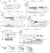
Fig. 3.. DTX3 monoubiquitylates TNKS on MAR.
(A) Schematic of DTX3 and DTX3L constructs. (B) Immunoblot analysis of TNKS1/2 DKO HEK293T cells transfected with the indicated plasmids, immunoprecipitated with anti-Flag antibody, stained with amido black, and probed with antibodies indicated on the left. (C) Immunoblot analysis of TNKS1/2 DKO HEK293T cells transfected with the indicated plasmids, immunoprecipitated with anti-Flag antibody, stained with amido black, and probed with antibodies indicated on the left. (D) Immunoblot analysis of TNKS1/2 DKO HEK293T cells transfected with the indicated plasmids, immunoprecipitated with anti-HA antibody, stained with amido black, and probed with antibodies indicated on the left. (E) Immunoblot analysis of TNKS1/2 DKO HEK293T cells transfected with the indicated plasmids, immunoprecipitated with anti-Flag antibody, stained with amido black, and probed with antibodies indicated on the left. (F) Immunoblot analysis of TNKS1/2 DKO HEK293T cells transfected with the indicated plasmids, immunoprecipitated with anti-Flag antibody, stained with amido black, and probed with antibodies indicated on the left. (G) Immunoblot analysis of TNKS1/2 DKO HEK293T cells transfected with the indicated Flag plasmids, immunoprecipitated with anti-HA antibody from denatured cell extracts, stained with amido black, and probed with antibodies on the left. (H) Immunoblot analysis of TNKS1/2 DKO HEK293T cells transfected with the indicated plasmids, immunoprecipitated with anti-HA antibody, stained with amido black, and probed with antibodies on the left. (I) Immunoblot analysis of TNKS1/2 DKO HEK293T cells transfected with the indicated plasmids, immunoprecipitated with anti-Flag antibody, stained with amido black, and probed with antibodies on the left. (J) Immunoblot analysis of TNKS1/2 DKO HEK293T cells transfected with the indicated plasmids, immunoprecipitated with anti-Myc antibody, stained with amido black, and probed with antibodies on the left. (K) Schematic showing DTX3 binds PAR and ubiquitylates MAR on TNKS. [(B), (E), (F), and (I)] Bacterial alkaline phosphatase (BAP) serves as a negative control.
Fig. 4.
Fig. 4.. Di19-UIM is a hybrid reader of the ubiquitin-MAR mark on TNKS.
(A) Schematic diagram showing MAR on protein. Sites of ubiquitin (Ub) addition and ADP-ribose (ADPr) addition are indicated. (B) Immunoblot analysis of TNKS1/2 DKO HEK293T cells transfected with the indicated plasmids, immunoprecipitated with anti-Flag antibody, stained with amido black, and probed with antibodies indicated on the left. Protein levels relative to TNKS and normalized to the control are indicated below the blots. (C) Schematic of RNF166 and Di19-UIM WT and mutant constructs. (D) Immunoblot analysis of TNKS1/2 DKO HEK293T cells transfected with the indicated plasmids, immunoprecipitated with anti-Flag antibody, stained with amido black, and probed with antibodies indicated on the left. (E) Graphical presentation of the change in MAR relative to TNKS1 and normalized to Empty Vector (EV). Average of four independent experiments ± SEM. **P ≤ 0.01, Student’s unpaired two-tailed t test. n.s., not significant. (F) Graphical presentation of the change in PAR relative to TNKS1 and normalized to EV. Average of four independent experiments ± SEM. **P ≤ 0.01, Student’s unpaired two-tailed t test. n.s., not significant. (G) Schematic showing the Di19-UIM hybrid reader binding to the MAR-Ub hybrid mark on TNKS. (H) Immunoblot analysis of TNKS1/2 DKO HEK293T cells transfected with the indicated plasmids, immunoprecipitated with anti-Flag antibody, stained with amido black, and probed with antibodies indicated on the left. (I) Graphical presentation of the change in MAR relative to TNKS1 and normalized to EV. Average of three independent experiments ± SEM. *P ≤ 0.05; **P ≤ 0.01, Student’s unpaired two-tailed t test. n.s., not significant. (J) Graphical presentation of the change in PAR relative to and normalized to EV. Average of three independent experiments ± SEM. *P ≤ 0.05; ***P ≤ 0.001, Student’s unpaired two-tailed t test. n.s., not significant.
Fig. 5.
Fig. 5.. Deltex and RINGUIM E3 ligases counter RNF146-mediated degradation of TNKS.
(A) Immunoblot analysis of extracts from TNKS1/2 DKO HEK293T cells transfected with FlagTNKS1 and the indicated Myc plasmids and probed with antibodies indicated on the left. Protein levels relative to tubulin and normalized to the control are indicated below the blots. (B) Immunoblot analysis of extracts from TNKS1/2 DKO HEK293T cells transfected with FlagTNKS1 and the indicated Myc plasmids and probed with antibodies indicated on the left. Protein levels relative to tubulin and normalized to the control are indicated below the blots. (C) Immunoblot analysis of extracts from TNKS1/2 DKO HEK293T cells transfected with FlagTNKS1 and the indicated Myc plasmids and probed with antibodies indicated on the left. Protein levels relative to tubulin and normalized to the control are indicated below the blots. (D) Immunoblot analysis of extracts from WT HEK293T cells transfected with the indicated Myc plasmids and probed with antibodies indicated on the left. Protein levels relative to tubulin and normalized to the control are indicated below the blots. (E) Graphical presentation of the change in TNKS relative to tubulin and normalized to control. Average of four independent experiments ± SEM. *P ≤ 0.05; **P ≤ 0.01; ****P ≤ 0.0001, Student’s unpaired two-tailed t test. (F) Immunoblot analysis of extracts from WT HEK293T cells transfected with the indicated Myc plasmids and probed with antibodies indicated on the left. Protein levels relative to tubulin and normalized to the control are indicated below the blots. (G) Graphical presentation of the change in TNKS relative to tubulin and normalized to control. Average of four independent experiments ± SEM. *P ≤ 0.05, Student’s unpaired two-tailed t test.
Fig. 6.
Fig. 6.. DTX3 is monoubiquitylated on MAR.
(A) Immunoblot analysis of TNKS1/2 DKO HEK293T cells transfected with the FLAGDTX3 or FLAGDTX3.RING** and HA-Ub, immunoprecipitated with anti-HA antibody, stained with amido black, and probed with antibodies indicated on the left. (B). Immunoblot analysis of TNKS1/2 DKO HEK293T cells transfected with the MycDTX3, HA-Ub, and the indicated Flag plasmids; immunoprecipitated with anti-HA antibody; stained with amido black; and probed with antibodies indicated on the left. (C) Immunoblot analysis of TNKS1/2 DKO HEK293T cells transfected with MycSAMPARP or MycDTX3, and FlagDi19-UIM, stained with amido black, and probed with antibodies indicated on the left. (*) indicates nonspecific band detected with the anti-MAR antibody. (D) Immunoblot analysis of TNKS1/2 DKO HEK293T cells transfected with the MycDTX3, HA-Ub, and the indicated Flag plasmids; immunoprecipitated with anti-HA antibody; stained with amido black; and probed with antibodies indicated on the left. (E) Immunoblot analysis of TNKS1/2 DKO HEK293T cells transfected with MycDTX3 and the indicated Flag plasmids, immunoprecipitated with anti-Myc antibody, stained with amido black, and probed with antibodies indicated on the left. (F) Immunoblot analysis of TNKS1/2 DKO HEK293T cells transfected with TNKS and the indicated Flag plasmids with and without MycDTX3, immunoprecipitated with anti-Flag antibody, stained with amido black, and probed with antibodies indicated on the left. (G) Schematic diagram showing a general mechanism where a MARylated protein target is monoubiquitylated on MAR by DTX2/3, followed by RNF114/166-mediated stabilization and dibuiquitylation. [(E) and (F)] Bacterial alkaline phosphatase (BAP) serves as a negative control.

References

    1. Luscher B., Ahel I., Altmeyer M., Ashworth A., Bai P., Chang P., Cohen M., Corda D., Dantzer F., Daugherty M. D., Dawson T. M., Dawson V. L., Deindl S., Fehr A. R., Feijs K. L. H., Filippov D. V., Gagne J. P., Grimaldi G., Guettler S., Hoch N. C., Hottiger M. O., Korn P., Kraus W. L., Ladurner A., Lehtio L., Leung A. K. L., Lord C. J., Mangerich A., Matic I., Matthews J., Moldovan G. L., Moss J., Natoli G., Nielsen M. L., Niepel M., Nolte F., Pascal J., Paschal B. M., Pawlowski K., Poirier G. G., Smith S., Timinszky G., Wang Z. Q., Yelamos J., Yu X., Zaja R., Ziegler M., ADP-ribosyltransferases, an update on function and nomenclature. FEBS J. 289, 7399–7410 (2021). - PMC - PubMed
    1. Suskiewicz M. J., Prokhorova E., Rack J. G. M., Ahel I., ADP-ribosylation from molecular mechanisms to therapeutic implications. Cell 186, 4475–4495 (2023). - PMC - PubMed
    1. Azarm K., Smith S., Nuclear PARPs and genome integrity. Genes Dev. 34, 285–301 (2020). - PMC - PubMed
    1. Jessop M., Broadway B. J., Miller K., Guettler S., Regulation of PARP1/2 and the tankyrases: Emerging parallels. Biochem. J. 481, 1097–1123 (2024). - PubMed
    1. Palazzo L., Mikoc A., Ahel I., ADP-ribosylation: New facets of an ancient modification. FEBS J. 284, 2932–2946 (2017). - PMC - PubMed

LinkOut - more resources