. 2025 Sep 5;11(36):eadx0005.
doi: 10.1126/sciadv.adx0005. Epub 2025 Sep 3.

Ndc80 complex, a conserved coupler for kinetochore-microtubule motility, is a sliding molecular clutch

Affiliations

Ndc80 complex, a conserved coupler for kinetochore-microtubule motility, is a sliding molecular clutch

Vladimir M Demidov et al. Sci Adv. .

Abstract

Chromosome motion at spindle microtubule plus ends relies on dynamic molecular bonds between kinetochores and proximal microtubule walls. Under opposing forces, kinetochores move bidirectionally along these walls while remaining near the ends, yet how continuous wall sliding occurs without end detachment remains unclear. Using ultrafast force-clamp spectroscopy, we show that single Ndc80 complexes, the primary microtubule-binding kinetochore component, exhibit processive, bidirectional sliding. Plus end-directed forces induce a mobile catch bond in Ndc80, increasing frictional resistance and restricting sliding toward the tip. Conversely, forces pulling Ndc80 away from the plus end trigger mobile slip-bond behavior, facilitating sliding. This dual behavior arises from force-dependent modulation of the Nuf2 calponin-homology domain's microtubule binding, identifying this subunit as a friction regulator. We propose that Ndc80's ability to modulate sliding friction provides the mechanistic basis for the kinetochore's end coupling, enabling its slip-clutch behavior.


Figures

Fig. 1.
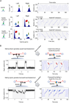
Fig. 1.. Bidirectional processive sliding of single Ndc80c molecules revealed by the UFFC assay.
(A) Kinetochores slide along the microtubules (MTs) wall in alternating directions, transitioning between the highly mobile “slip” state and the “clutch” state, which generates higher molecular friction to protect the trailing kinetochore from detachment at the microtubule plus end. (B) Ndc80c binds microtubule wall with the toe of the Hec1 calponin-homology domain (CHD) with a pronounced tilt toward the microtubule plus end. (C) Schematic of the three-bead assay setup and a representative image showing merged fluorescence and differential interference contrast channels. (D) Representative signals showing changes in the position of a dumbbell bead (blue) and the clamped force (red) during experiments with GFP- or Ndc80c-coated pedestals. Gray area corresponds to a continuous gliding interaction.
Fig. 2.
Fig. 2.. Sliding friction of single Ndc80c molecules is strongly microtubule-polarity dependent.
(A) Histograms showing distributions of instantaneous velocities in two oscillation directions along with typical coordinate recordings of the dumbbell beads for 30 s at 4-pN clamp force. Rightward dumbbell oscillation corresponds to an upward change in bead coordinate on the graphs, while leftward dumbbell motion results in a decreasing bead coordinate. The first row depicts a control experiment with GFP-coated pedestals, showing free velocity peaks in two directions (green arrows). The following rows show results from two different dumbbells oscillated near Ndc80c-coated pedestals. Red and blue arrows indicate slower than normal velocity peaks, which were observed in different oscillation directions in these two examples. (B) Changes in the coordinate of the dumbbell beads during motor pulling or trap-induced Ndc80c sliding, tested using the same microtubule dumbbells.
Fig. 3.
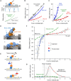
Fig. 3.. Analysis of force-velocity relationship.
(A) Schematic of Ndc80c diffusing in a periodic, single-site energy landscape, where microtubule binding is mediated by the Hec1 CHD. Sliding velocity is expected to increase exponentially with applied force. At low force, velocity is set by the intrinsic rate of force-free diffusion D, which determines the slope (40). (B) Ndc80c velocity under a clamped force applied to the dumbbell, dragging Ndc80c along a microtubule. Each colored point represents mean velocity during unidirectional sweeps (average of 2042 sweeps per point) from 30-s recordings (n = 106, N = 43 chambers). Curves show exponential fits. Open symbols indicate dumbbell oscillation velocity without Ndc80c binding; lower Ndc80c velocity reflects stronger friction. (C) Velocities from (B) plotted against forces acting directly on Ndc80c. Dark circles show binned data (means ± SD). Both curves deviate from the single-site model (green) with D = 0.11 μm2/s and a 9-kBT well, as in force-free Ndc80c diffusion (32). (D) Docking of crystal structures for Hec1 and Nuf2 CHDs on a microtubule wall segment. The Ndc80c stalk and other regions were omitted. (E) Two-site model. Arrows represent diffusional steps via the Hec1 CHD toe binding. Plus end–directed forces promote Nuf2 CHD tubulin contact, increasing binding energy; minus end–directed forces pull Nuf2 CHD away. (F) Solution of the two-site Brownian model with 6-kBT well depths for both binding sites and a rotational stiffness of 10−2 pN·μm. The black line is a polynomial fit through the origin. Experimental data (pale circles) and the single-site model (green) are from (C). Negative forces correspond to minus end–directed pulling. (G) Friction coefficient of Ndc80c (black line). Gray shading indicates the force range where friction rises steeply. The single-site model fails to capture this complex dependence despite showing similar behavior at zero force.
Fig. 4.
Fig. 4.. Nuf2 CHD, but not Hec1 tail, is required for friction generation.
(A) Crystal structure of the Hec1 and Nuf2 CHDs [Protein Data Bank (PDB) ID 2VE7] (25) showing mutated residues (red) and the not-to-scale drawing of the unstructured Hec1 N-terminal extension. (B) Example bead coordinate signals in experiments using the indicated Ndc80c Bonsai proteins at 4-pN clamp force. Tandem slow velocity segments (highlighted with light gray) are evident for all proteins, but sliding is faster in CHD mutants. (C) Force-velocity dependencies for the plus end–directed sliding determined with UFFC. Each symbol shows mean and SD for different force bins based on N = 43 chambers for WT, N = 6 for ΔTail, N = 12 for 3D Nuf2, and N = 8 for Hec1 K166D. (D) Images of rhodamine-labeled taxol-stabilized microtubules and their decoration by the indicated GFP-tagged Ndc80c Bonsai proteins (100 nM). Image with unmodified Bonsai Ndc80c-GFP has reduced contrast relative to other GFP images because of its excessive brightness. (E) Brightness of microtubule decoration with indicated GFP-tagged Ndc80c proteins plotted on the semilog scale. Each dot shows average GFP fluorescence of one microtubule, and bars and whiskers show means ± SD. a.u., arbitrary units.

Update of

References

    1. Walczak C. E., Cai S., Khodjakov A., Mechanisms of chromosome behaviour during mitosis. Nat. Rev. Mol. Cell Biol. 11, 91–102 (2010). - PMC - PubMed
    1. Vladimirou E., Harry E., Burroughs N., McAinsh A. D., Springs, clutches and motors: Driving forward kinetochore mechanism by modelling. Chromosome Res. 19, 409–421 (2011). - PMC - PubMed
    1. Monda J. K., Cheeseman I. M., The kinetochore–microtubule interface at a glance. J. Cell Sci. 131, jcs214577 (2018). - PMC - PubMed
    1. Skibbens R., Skeen V., Salmon E., Directional instability of kinetochore motility during chromosome congression and segregation in mitotic newt lung cells: A push-pull mechanism. J. Cell Biol. 122, 859–875 (1993). - PMC - PubMed
    1. Skibbens R. V., Rieder C. L., Salmon E. D., Kinetochore motility after severing between sister centromeres using laser microsurgery: Evidence that kinetochore directional instability and position is regulated by tension. J. Cell Sci. 108 ( Pt. 7), 2537–2548 (1995). - PubMed

LinkOut - more resources