. 2025 Aug 27;53(16):gkaf843.
doi: 10.1093/nar/gkaf843.

The endoribonuclease Rae1 from Bacillus subtilis cleaves mRNA upstream of stalled ribosomes

Affiliations

The endoribonuclease Rae1 from Bacillus subtilis cleaves mRNA upstream of stalled ribosomes

Valentin Deves et al. Nucleic Acids Res. .

Abstract

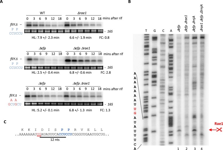
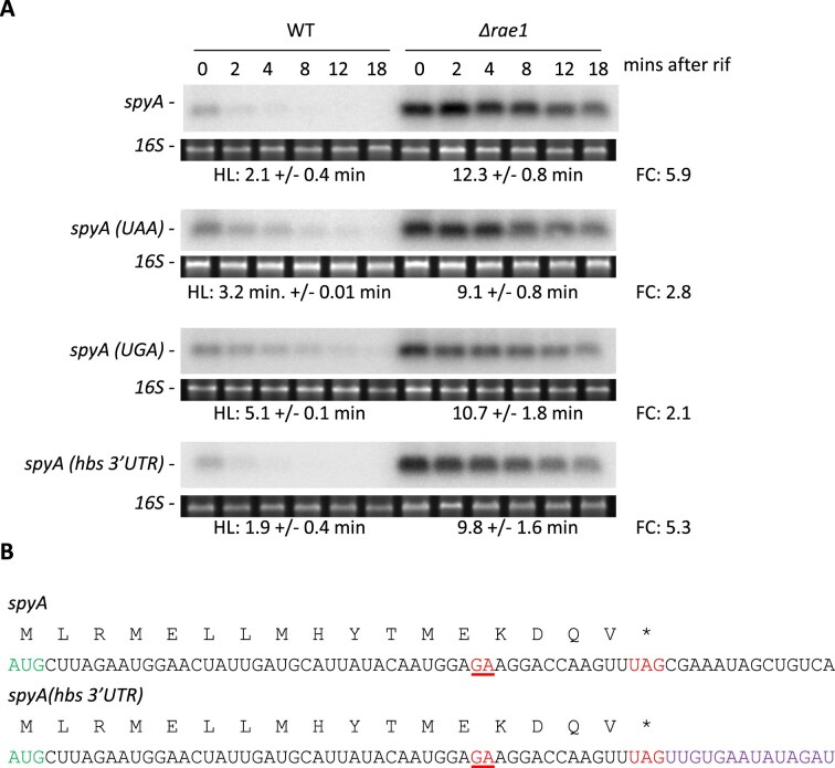
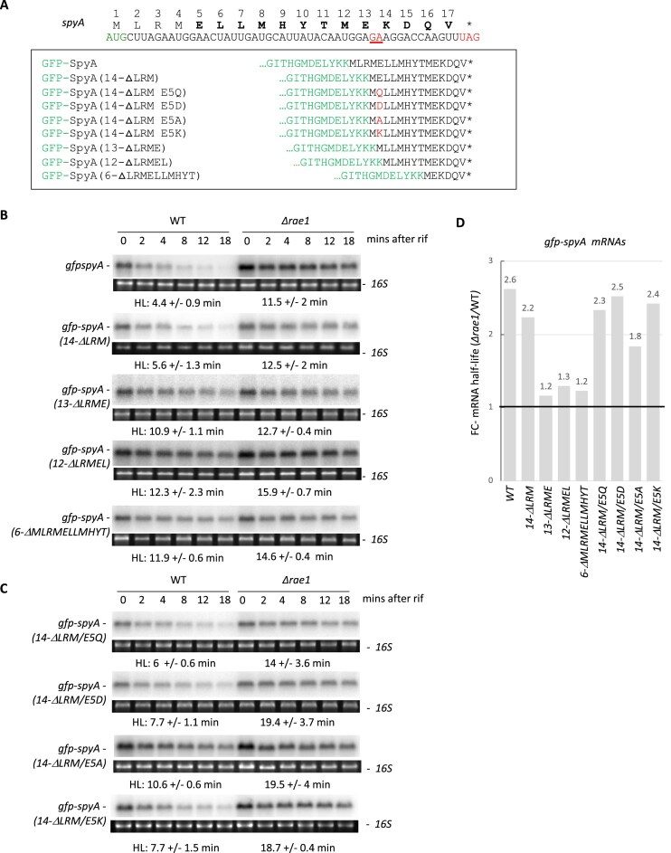
The ribosome-associated endoribonuclease 1 (Rae1) cleaves messenger RNAs (mRNAs) in a translation-dependent manner. Here, we identify a new Rae1 target, the fliY mRNA, which is cleaved by Rae1 in the absence of the elongation factor P. The Rae1 site was mapped 12 nucleotides upstream of the second proline codon of an SPP stalling motif in fliY. Remarkably, Rae1 cleavages also occur 12 nucleotides upstream of the stop codon within two validated Rae1 mRNA targets, bmrX and spyA (S1025). Shifting the stop codon relative to the Rae1 cutting site abolished Rae1 sensitivity of bmrX and spyA mRNAs. We show that ribosome pausing occurs at the spyA stop codon, confirming its crucial role, and positioning the Rae1 cleavage at the tail end of the stalled ribosome, rather than in the A-site as previously proposed. These findings reveal a compelling novel mechanism by which Rae1 mediates mRNA cleavage in coordination with immobile ribosomes.


Conflict of interest statement

None declared.

Figures

Graphical Abstract
Graphical Abstract
Figure 1.
Figure 1.
Rae1 cleaves the fliY mRNA upstream of the SPP stalling motif. (A) Stability of fliY mRNA revealed by northern blot. The stability of the truncated version of fliY(fliYΔ) and its derivative with the SPP motif mutated to SAA [fliYΔ (P165A P166A)], inserted into the chromosome at the amyE locus, was analyzed in WT, Δrae1, Δefp, and ΔefpΔrae1 strains. RNAs were extracted 0, 3, 6, 9, 12, and 18 min after addition of rifampicin at 150 μg/ml. The blots were probed with oligonucleotide CC2840. HLs were calculated from biological replicates. The fold change (FC) in HLs is given next to each autoradiogram. (B) Cleavage site of fliYΔ mRNA by Rae1 determined by primer extension assay. Total RNA from Δefp, ΔefpΔrae1, ΔefpΔrnjA, and ΔefpΔrae1ΔrnjA strains containing the fliYΔ gene inserted at amyE were subject to reverse transcription. Primer extension was performed with oligonucleotide CC2947. The Rae1 cleavage site is highlighted in red. (C) Localization of Rae1 cleavage site on the fliY mRNA. The Rae1 cleavage site (underlined in red) is located 12 nt upstream of the second proline codon of the SPP stalling motif (in bold). The 12-nt distance between the proline codon and the Rae1 cleavage site is represented by a black line.
Figure 2.
Figure 2.
Ribosomes stall at the spyA stop codon. The in vitro-transcribed WT or F+1 spyA mRNAs were translated in vitro and toe-print assays performed by primer extension using primer CC1661. RNAs were incubated without (−) (lanes 1 and 5) or with 2.7 pmol of B. subtilis ribosomes (+) (lanes 2–4 and 6–8), in the presence of 100 μg/ml of tetracycline (lanes 3 and 7) or 100 μg/ml of puromycin (lanes 4 and 8). Blue arrow: toe-print at +17 from start site. Orange arrow: toe-print at +17 from termination site. The sequences of WT and F+1 spyA are shown in the lower panel. The modified nucleotides in the F+1 spyA sequence, insertion of a C and substitution of U6C, are indicated in purple. Green: start codon. Red: stop codon. Blue and orange: toeprint signals. Red underlined: Rae1 cleavage site.
Figure 3.
Figure 3.
The distance between the Rae1 cleavage site and the stop codon is important for Rae1 cleavage (A) Rae1 cleavage sites in the bmrX and spyA mRNAs. The Rae1 cleavage site is underlined in red. The nucleotide distance between the stop codon and the Rae1 site is represented by a black line. Start codons are highlighted in green, stop codons in red and the 18-nt random sequence added downstream of spyA mRNA in blue [spyA(+18 nts)]. In the spyA UAG(+18 nts) construct, a stop codon was reintroduced at its original location. (B) Stability of various spyA transcripts in WT and Δrae1 strains revealed by northern blot. Total RNA extracted 0, 2, 4, 8, 12, and 18 min after addition of rifampicin at 150 μg/ml. The blots were probed with oligonucleotide CC2799 that hybridized to spyA. The HLs of spyA, spyA(+18 nts), and spyA UAG(+18 nts) were calculated from two biological replicates and are given under each autoradiogram. The FC in HLs is given next to each blot. (CIn vitro cleavage assay of spyA by Rae1 during translation. One pmol of 5′- labeled spyA, spyA(+18 nts), and spyA UAG(+18 nts) transcripts were incubated in presence of 2.7 pmol of ribosomes and 38 pmol of Rae1. A control for RNA integrity was added without ribosomes or Rae1. The 72-nt band corresponding to the cleaved transcript is highlighted in red.
Figure 4.
Figure 4.
The stop codon plays a crucial role in Rae1 cleavage of spyA (A) Sequence of spyA-gfp translational fusion. The gfp coding sequence was fused upstream (gfp-spyA) or downstream (spyA-gfp) of the spyA ORF. A stop codon was placed in the spyA(UAG)-gfp fusion 12 nt from the Rae1 site (underlined in red). Black: spyA sequence. Green: gfp sequence. Bold green: start codon. Bold red: stop codon. (B) Stability of mRNA spyA-gfp translational fusion revealed by northern blot in WT and Δrae1 strains. RNAs were extracted 0, 2, 4, 8, 12, and 18 min after addition of rifampicin at 150 μg/ml. HLs were calculated from two biological replicates. The blots were probed with oligonucleotide CC2422 that hybridized to gfp. The FC in HLs is given next to each autoradiogram.
Figure 5.
Figure 5.
The nature of the stop codon impacts Rae1 cleavage efficiency (A) Stability of the spyA mRNA with different stop codons or a different 3′-UTR revealed by northern blots in WT and Δrae1 strains. Strains expressing the spyA mRNA sequence carrying either UAG, UAA, or UGA stop codons, or the hbs 3′-UTR, were generated. RNAs were extracted 0, 2, 4, 8, 12, and 18 min after addition of rifampicin at 150 μg/ml. The blots were probed with CC2799. HLs were calculated from two biological replicates. The FC in HLs is given next to each autoradiogram. (B) In the spyA(hbs3’UTR) construct, the 3′UTR of spyA was replaced by the hbs 3′-UTR (in violet).
Figure 6.
Figure 6.
Effects of RF1 and RF2 on sensitivity to Rae1 cleavage. In vitro cleavage assay of spyA by Rae1 in the presence of RF1 or RF2. One pmol of the spyA in vitro transcript, recognized by RF1, or spyA(UGA), recognized by RF2, was incubated in presence of 2.7 pmol of ribosomes alone (lanes 1 and 7), 38 pmol of Rae1 (lanes 3 and 9), or with 2 pmol (lanes 4 and 10) or 20 pmol (lanes 5–6 and 11–12) of B. subtilis RF1 or RF2. The band corresponding to the transcript cleaved by Rae1 is highlighted in red. The percentage of the 72-nt cleavage product was determined from three independent experiments. The FC in the presence of the RF factor compared to its absence, is shown below the lanes [a P-value of <.05 is indicated by an asterisk (*)].
Figure 7.
Figure 7.
Modification of the SpyA or BmrX peptide sequence inhibits Rae1 cleavage. (A) The sequences of spyA and bmrX were mutated to modify the peptide sequence without altering the 12-nt distance between the Rae1 cleavage site and the stop codon. The mutated nucleotides are shown in purple as well as the modified amino acids. Green: start codon. Red: stop codon. Red underlined: Rae1 cleavage site. (B) Stability of the spyA-AAmod and bmrX-AAmod mRNAs, respectively, revealed by northern blot in the WT and Δrae1 strains. RNAs were extracted 0, 2, 4, 8, 12, and 18 min after addition of rifampicin at 150 μg/ml. The blots were probed with CC2799 or CC3271. HLs were calculated from two biological replicates. The FC in HLs is given next to each autoradiogram. (CIn vitro cleavage assay of spyA and spyA-AAmod mRNAs by Rae1 during translation. One pmol of 5′-labeled mRNAs was incubated in the presence of 2.7 pmol of ribosomes and 38 pmol of Rae1. A control for RNA integrity was added without ribosomes or Rae1. The band corresponding to the transcript cleaved by Rae1 is highlighted in red.
Figure 8.
Figure 8.
Shortening the spyA ORF makes the transcript insensitive to Rae1. (A) Peptide and nucleotide sequence of spyA. The Rae1 cleavage site is underlined in red, the start codon in green, and the stop codon in red. (B) Stability of spyA mRNAs of various lengths in WT and Δrae1 strains revealed by northern blot. RNAs were extracted 0, 2, 4, 8, 12, and 18 min after addition of rifampicin at 150 μg/ml. HLs were calculated using two biological replicates. The blots were probed with oligonucleotide CC2799. The spyA sequence (17 codons) was reduced by serial deletion from the N-terminus of 1 codon (16-ΔL), 2 codons (15-ΔLR), or 3 codons (14-ΔLRM), or by deletion of codons 5–7 (14-ΔELL). The LRM amino acids from position 2 to 4 were also substituted by FKV (17-FKV) or RML (17-RML). (C) FC of mRNA HLs between Δrae1 and WT strains for the different spyA constructs. (DIn vitro translation cleavage assay of spyA and spyA(14-ΔLRM) in the presence of 2.7 pmol ribosomes and 38 pmol Rae1. A control for RNA integrity was added without ribosomes and Rae1. The band corresponding to the transcript cleaved by Rae1 is highlighted in red.
Figure 9.
Figure 9.
The 5ELLMHYTMEKDQV17 sequence is essential for Rae1-induced destabilization of spyA. (A) Design of gfp-spyA translational fusions. Peptide and nucleotide sequence of spyA with the Rae1 cleavage site underlined in red, start codon in green, and stop codon in red. The WT 17-codon sequence or shorter versions were cloned in frame downstream of the gfp ORF (green) and containing, respectively, the last 14 codons (14-ΔLRM), 13 codons (13-ΔLRME), 12 codons (12-ΔLRMEL), or 6 codons (6-ΔMLRMELLMHYT) of spyA. The role of glutamate at position 5 was investigated by replacing the E5 residue with an aspartate [gfp-spyA(14-ΔLRM/E5D)], glutamine [gfp-spyA(14-ΔLRM/E5Q)], alanine [gfp-spyA(14-ΔLRM/E5A)], or lysine [gfp-spyA(14-ΔLRM/E5K)]. (B and C) Stability of the gfp-spyA translational fusions with shortened spyA moieties in WT and Δrae1 strains revealed by northern blot. RNAs were extracted 0, 2, 4, 8, 12, and 18 min after addition of rifampicin at 150 μg/ml. HLs were calculated from biological replicates. The blots were probed with oligonucleotide CC2422. (D) FC of mRNA HLs between Δrae1 and WT strains for the different spyA constructs.

References

    1. Deana A, Belasco JG Lost in translation: the influence of ribosomes on bacterial mRNA decay. Genes Dev. 2005; 19:2526–33. 10.1101/gad.1348805. - DOI - PubMed
    1. Durand S, Tomasini A, Braun F et al. sRNA and mRNA turnover in Gram-positive bacteria. FEMS Microbiol Rev. 2015; 39:316–30. 10.1093/femsre/fuv007. - DOI - PubMed
    1. Radhakrishnan A, Green R Connections underlying translation and mRNA stability. J Mol Biol. 2016; 428:3558–64. 10.1016/j.jmb.2016.05.025. - DOI - PubMed
    1. Trinquier A, Durand S, Braun F et al. Regulation of RNA processing and degradation in bacteria. Biochimica Et Biophysica Acta Gene Regul Mech. 2020; 1863:194505. 10.1016/j.bbagrm.2020.194505. - DOI - PubMed
    1. Bae H, Coller J Codon optimality-mediated mRNA degradation: linking translational elongation to mRNA stability. Mol Cell. 2022; 82:1467–76. 10.1016/j.molcel.2022.03.032. - DOI - PMC - PubMed

MeSH terms

LinkOut - more resources