. 2025 Dec;17(1):2547029.
doi: 10.1080/19490976.2025.2547029. Epub 2025 Sep 3.

Gut microbiota dysbiosis affects intestinal sensitivity through epithelium-to-neuron signaling: novel insights from a colon organoid-based model to improve visceral pain therapy

Affiliations

Gut microbiota dysbiosis affects intestinal sensitivity through epithelium-to-neuron signaling: novel insights from a colon organoid-based model to improve visceral pain therapy

Francesco Margiotta et al. Gut Microbes. 2025 Dec.

Abstract

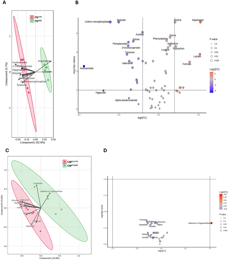
Chronic gastrointestinal pain is a hallmark of most intestinal pathologies, yet effective treatments remain elusive given the complexity of the underlying mechanisms. Aiming to investigate the intestinal epithelium contribution to visceral pain modulation in dysbiosis context, we first demonstrated that intracolonic instillation of microbe-free fecal supernatants from mice with post-inflammatory dysbiosis induced by dextran sodium sulfate (FSDSS) provokes visceral hypersensitivity in recipient mice. Epithelium involvement in the response to FSDSS was analyzed through a novel in vitro approach comprising murine epithelial colon organoids and primary dorsal root ganglia (DRG) neurons. FSDSS treatment induced growth and metabolic impairment in colon organoids, which revealed a dysbiosis-driven epithelial dysfunction. Notably, the combination of FSDSS and conditioned medium from FSDSS-treated colon organoids induced an increase in DRG neuron intrinsic excitability, along with greater immunoreactivity to c-Fos and calcitonin-gene related peptide, implicating an integrated role of both microbial and epithelial products in visceral sensitivity regulation. By investigating the underlying signaling, metabolomic analysis revealed reduced levels of short chain fatty acids in FSDSS, such as butyrate, acetate, valerate, and propionate. Moreover, transcriptomic analysis of FSDSS-treated colon organoids showed the dysregulated expression of several signaling factors by which intestinal epithelium may modulate sensory neuron excitability, including proteases, cytokines, neuromodulators, growth factors, and hormones. These findings provide novel insights into the role of gut epithelium in the modulation of sensory neuron excitability under dysbiosis conditions, emphasizing that targeting epithelial-neuronal signaling might represent a promising therapeutic strategy for visceral pain management.

Keywords: DRG neurons; Visceral pain; dysbiosis; epithelial-neuronal signaling; intestinal epithelium; microbiota; organoids.


Conflict of interest statement

No potential conflict of interest was reported by the author(s).

Figures

Figure 1.
Figure 1.
Effects of FSCTR and FSDSS intracolonic injection on visceral sensitivity of naïve mice - assessment of abdominal withdrawal reflex to colorectal distension. A) FSCTR and FSDSS (300 µL 100 mg/mL) were injected in naïve animals once daily for 4 consecutive days and AWR was assessed 1 and 24 hours after the first FS injection, 3 and 7 days after the last FS injection, as reported in the scheme. Visceral sensitivity was measured by evaluating the AWR (score 0–4) in response to colorectal distension (50–200 µL) B) 1 and C) 24 hours after the first FS injection, D) 3 and E) 7 days after the last FS injection. Values represent the mean ± SEM of each experimental group. The analysis of variance was performed by one-way ANOVA followed by Bonferroni post hoc comparison.
Figure 2.
Figure 2.
Effects of FSCTR and FSDSS on viability and growth ability of colon organoids. A) Colon organoids were treated on days 3 and 5 from the seeding (day 1) with 4 mg/mL FSCTR and FSDSS, and experimental analysis were performed on day 7. B) MTT assay was performed on colon organoids following FS treatments to evaluate the effects of FS on their viability. C) Morphometric analysis was performed on colon organoids following FS treatments to assess the impact of FS on their growth ability. D) Representative images of organoids after treatment with FS (magnification: 20×; scale bar: 100 µm). Values represent the mean ± SEM of n=3 experiments for MTT assay and n=5 experiments for morphometric analysis. The analysis of variance was performed by one-way ANOVA followed by Bonferroni post hoc comparison.
Figure 3.
Figure 3.
Electrophysiological analysis on DRG neurons exposed to FS and/or CM from colon organoids. A) DRG neurons were exposed for 48 hours to CMCTR, FSCTR, FSDSS, CMFS CTR, CMFS DSS, CMFS CTR + FSCTR and CMFS DSS + FSDSS. Intrinsic excitability was measured in B) FSCTR vs FSDSS, C) CMFS CTR vs CMFS DSS and D) CMFS CTR + FSCTR vs CMFS DSS + FSDSS. Example traces of action potentials recordings were obtained in response to a +125 pA current. Values represent the mean ± SEM of 6–12 cells analyzed in n=4 experiments. The analysis of variance was performed by one-way ANOVA followed by Bonferroni post hoc comparison.
Figure 4.
Figure 4.
Immunofluorescence analysis for c-Fos and CGRP in DRG neurons exposed to FS and/or CM from colon organoids A) Representative images of DRG neurons stained for c-Fos (green) and CGRP (red) after 48 hours exposure to CMFS CTR + FSCTR and CMFS DSS + FSDSS (magnification: 40×; scale bar: 100 µm). Fluorescence intensity was measured for B) c-Fos and C) CGRP in all experimental groups. Values represent the mean ± SEM of n=4–6 different slides for each condition. The analysis of variance was performed by one-way ANOVA followed by Bonferroni post hoc comparison.
Figure 5.
Figure 5.
Assessment of c-Fos expression in different subpopulations of DRG neurons exposed to CMFS CTR + FSCTR and CMFS DSS + FSDSS. Four subpopulations of DRG neurons with different proportions were identified in A) CMFS CTR + FSCTR and B) CMFS DSS + FSDSS groups, based on IB4-binding affinity and CGRP expression. C) The immunoreactivity for c-Fos was measured in IB4+, CGRP+, CGRP+/IB4+, and CGRP/IB4 subpopulations comparing the two experimental conditions. Lines represent the median within the box, the 25th and 75th percentiles at the ends of the box (interquartile range), and the error bars define the 25th + 1.5 interquartile range and the 75th + 1.5 interquartile range. The analysis of variance was performed by Kruskal-Wallis test followed by Dunn post hoc comparison.
Figure 6.
Figure 6.
Analysis of colon organoid transcriptomic profile under FSCTR and FSDSS treatments. A) Volcano plot shows the genes that are differentially expressed in organoids in the comparison FSDSS vs FSCTR. Gene set enrichment analysis (GSEA) was performed in B) Hallmark and C) GO:BP datasets to identify which pathways emerge in the comparison FSDSS vs FSCTR. RNA sequencing analysis was conducted on n=4 samples for each condition. Genes with p<0.05 and Log2(FC)≥0.58 or ≤-0.58 were considered as significant DEGs. Pathways significantly modulated were selected according to the adjusted p<0.05.
Figure 7.
Figure 7.
Expression of relevant genes in colon organoids under FSCTR and FSDSS treatments. The analysis was conducted for genes coding for A) serine proteases and pappalysins, B) ADAMs and ADAMTS, C) cathepsins and calpains, and D) neuromodulators. Genes in the red area were upregulated in FSDSS while genes in the blue area were downregulated in FSDSS.
Figure 8.
Figure 8.
Metabolomic characterization of fecal supernatants and conditioned media from organoids. Metabolomic differences in FSDSS vs FSCTR and CMFS DSS vs CMFS CTR comparisons were reported respectively in A, C) PLS-DA and B, D) volcano plots. The analysis was conducted on n=5 samples for each condition. Adjusted p<0.1 was considered statistically significant.

References

    1. Drossman DA. Functional gastrointestinal disorders: history, pathophysiology, clinical features, and Rome IV. Gastroenterology. 2016;150:1262–1279.e2. doi: 10.1053/j.gastro.2016.02.032. - DOI - PubMed
    1. Ford AC, Vanner S, Kashyap PC, Nasser Y. Chronic visceral pain: new peripheral mechanistic insights and resulting treatments. Gastroenterology. 2024;166:976–26. doi: 10.1053/j.gastro.2024.01.045. - DOI - PMC - PubMed
    1. Sinopoulou V, Gordon M, Dovey TM, Akobeng AK. Interventions for the management of abdominal pain in ulcerative colitis. Cochrane Database Syst Rev. 2021;CD013589. doi: 10.1002/14651858.CD013589.pub2. - DOI - PMC - PubMed
    1. Grundy L, Erickson A, Brierley SM. Visceral pain. Annu Rev Physiol. 2019;81:261–284. doi: 10.1146/annurev-physiol-020518-114525. - DOI - PubMed
    1. Lucarini E, Di Pilato V, Parisio C, Micheli L, Toti A, Pacini A, Visceral sensitivity modulation by faecal microbiota transplantation: the active role of gut bacteria in pain persistence. Pain. 2022;163:861–877. doi: 10.1097/j.pain.0000000000002438. - DOI - PMC - PubMed

MeSH terms

LinkOut - more resources