. 2025 Sep;31(9):e70578.
doi: 10.1111/cns.70578.

Endothelin-Converting Enzyme-Like 1 Regulated by LIF Contributes to Chronic Constriction Injury-Induced Neuropathic Pain in Mice

Affiliations

Endothelin-Converting Enzyme-Like 1 Regulated by LIF Contributes to Chronic Constriction Injury-Induced Neuropathic Pain in Mice

Feng Gao et al. CNS Neurosci Ther. 2025 Sep.

Abstract

Aims: This study is to investigate the role of Endothelin-converting enzyme-like 1 (ECEL1) in neuropathic pain (NP).

Methods: The expression of ECEL1 was modulated by injecting adeno-associated virus 5 (AAV5) carrying Ecel1 shRNA or full-length Ecel1 into the dorsal root ganglion (DRG) of mice with a chronic constriction injury (CCI) model. Then, various nociceptive responses were evaluated. Additionally, leukemia inhibitory factor (LIF) was intrathecally injected, or its function was blocked, to observe the changes in ECEL1 expression.

Results: Our findings demonstrate that downregulating ECEL1 expression alleviates CCI-induced pain and reduces the hyperexcitability of injured DRG neurons, which is achieved by inhibiting sympathetic sprouting in the DRG. Conversely, overexpressing ECEL1 in DRG neurons leads to pain hypersensitivity. Additionally, we observed that LIF upregulated ECEL1 expression, while blocking LIF reduced ECEL1 expression and mitigated CCI-induced nociception in mice.

Conclusion: ECEL1 promotes hyperalgesia following CCI and is regulated by LIF, suggesting it could be a new target for NP treatment.

Keywords: DRG; ECEL1; LIF; neuropathic pain; sympathetic sprouting.


Conflict of interest statement

The authors declare no conflicts of interest.

Figures
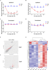
FIGURE 1
FIGURE 1
Analysis of DEGs in the DRG in the context of neuropathic pain induced by CCI. (A–D) Assessment of paw withdrawal responses to mechanical stimuli (A, C) and thermal stimuli (B, D) on the ipsilateral and contralateral sides of mice subjected to CCI compared with those in the sham group. n = 6 per group. (E) Upregulated and downregulated genes in the ipsilateral L4 and L5 DRGs in the CCI group compared with the sham group. (F) Volcano plot showing the upregulated and downregulated genes in the ipsilateral L4 and L5 DRGs in the CCI group compared with the sham group. The red dots represent genes whose expression was significantly upregulated, the blue dots represent genes whose expression was significantly downregulated, and the gray dots represent genes whose expression was not significantly different. (G) Heatmap showing the hierarchical clustering of DEGs in the CCI group compared with the sham group. The results are expressed as the mean ± SEM; *p < 0.05, **p < 0.01, ***p < 0.001 versus the sham group. Two‐way ANOVA with Tukey's post hoc test was used in A–D.
FIGURE 2
FIGURE 2
Expression and cellular distributions of ECEL1 in the DRG in mice after CCI. (A, B) Levels of Ecel1 mRNA in the ipsilateral and contralateral L4 and L5 DRGs after sham (A) or CCI (B) surgery. n = 6 per group. (C, E) Western blots (C) and statistical analysis (E) of ECEL1 protein levels in the ipsilateral L4 and L5 DRGs after sham surgery. (D, F) Western blots (D) and statistical analysis (F) of ECEL1 protein levels in the ipsilateral L4 and L5 DRGs after CCI. n = 6 per group. The results are expressed as the mean ± SEM; *p < 0.05, **p < 0.01, ***p < 0.001 versus the control group. Two‐way ANOVA with Tukey's post hoc test was used in A and B. One‐way ANOVA with Tukey's post hoc test was used in E and F.
FIGURE 3
FIGURE 3
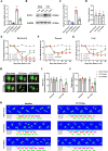
DRG ECEL1 knockdown attenuates CCI injury‐induced nociception development in mice. (A) Levels of Ecel1 mRNA in the L4 and L5 DRGs of mice that were injected with shRNA or scrambled shRNA on day 21 after sham or CCI surgery. (B, C) Western blots (B) and statistical analysis (C) of ECEL1 in the L4 and L5 DRGs of each group of mice. (D) Locomotor performance in the rotarod test of different groups of mice on day 21 after sham or CCI surgery. (E–G) Effects of microinjection of shRNA or Scram into the ipsilateral L4 and L5 DRGs on paw withdrawal responses to mechanical (E), thermal (F), and cold (G) stimuli on the ipsilateral side on the indicated days before or after CCI surgery in mice. (I and J) Percentages of the ipsilateral paw print area (I) and single stance (J) assessed by CatWalk analysis (% ipsilateral/contralateral). (H) Combined paw print image. (K) Representative digitized paw prints and associated step cycles. The results are expressed as the means ± SEMs; *p < 0.05, **p < 0.01, ***p < 0.001 versus the sham + Scram group; # p < 0.05, ## p < 0.01, ### p < 0.001 versus the CCI + Scram group. One‐way ANOVA with Tukey's post hoc test was used in A, C, and D. Two‐way ANOVA with Tukey's post hoc test was used in E‐G, I, and J. n = 6 per group.
FIGURE 4
FIGURE 4
Specific downregulation of ECEL1 alters the excitability of DRG neurons in CCI mice. (A) Current–clamp recordings of the action potential traces of GFP‐positive neurons in DRGs collected from CCI mice 2 weeks after the injection of shRNA or Scram. (B) Quantification of the firing frequency of action potentials in GFP‐positive neurons in the DRG in CCI mice injected with Ecel1 shRNA and Scram. (C–E) Quantification of the amplitude (C), resting membrane potential (D), and threshold of action potential generation (E) in GFP‐positive DRG neurons from different groups of mice. The results are expressed as the means ± SEMs; *p < 0.05, **p < 0.01, ***p < 0.001 versus the Sham + Scram group; #p < 0.05, ##p < 0.01 versus the CCI + Scram group. Two‐way ANOVA with Tukey's post hoc test was used in B. One‐way ANOVA with Tukey's post hoc test was used in C–E; n = 12 per group.
FIGURE 5
FIGURE 5
ECEL1 overexpression in sensory neurons of the DRG evokes hyperalgesia symptoms in naive mice. (A–C) Levels of Ecel1 mRNA (A) and protein (B, C) in the ipsilateral L4 and L5 DRGs after microinjection of an AAV5‐Ecel1‐overexpressing virus or an AAV5‐GFP control virus into the unilateral L4 and L5 DRGs in mice. N = 6 per group. (D–F) Paw withdrawal responses to mechanical (D), thermal €, and cold (F) stimuli on the ipsilateral side after microinjection of an ECEL1‐overexpressing strain and a GFP virus into the ipsilateral L4 and L5 DRGs in naive mice. n = 6 per group. (H) Representative images showing the basket structures (red) around DRG neurons in each group of mice (scale bar, 50 μm). (G) Statistical analysis of the percentage of DRG neurons surrounded by TH‐IR baskets. n = 12 per group. The results are expressed as the mean ± SEM; *p < 0.05, **p < 0.01, ***p < 0.001 versus the sham or control group, ### p < 0.001 versus the CCI + Scram group. Student's t test was used in A, C. Two‐way ANOVA with Tukey's post hoc test was used in D–F. One‐way ANOVA with Tukey's post hoc test was used in G.
FIGURE 6
FIGURE 6
ECEL1 is regulated by LIF. (A, B) Western blots (A) and statistical analysis (B) of LIF protein levels in the DRG after CCI. n = 6 per group. (C, D) Western blots (C) and statistical analysis (D) of the expression of ECEL1 in the DRG in mice injected with gp130 shRNA or Scram 14 days before sham or CCI surgery. (E–G) ECEL1 protein levels (E, F) and quantitative PCR assays of ECEL1 mRNA levels (G) in the DRG after the intrathecal injection of LIF. n = 6 per group. (H–K) Paw withdrawal responses to mechanical (H, I) and thermal (J, K) stimuli on the ipsilateral side and contralateral side in CCI or sham mice injected with gp130 shRNA or Scram. n = 6 per group. The results are expressed as the mean ± SEM; ***p < 0.005 versus the sham or control group; ## p < 0.01 and ### p < 0.001 versus the CCI + Scram group. Student's t test was used in B, F, G. One‐way ANOVA with Tukey's post hoc test was used in D. Two‐way ANOVA with Tukey's post hoc test was used in H–K.
FIGURE 7
FIGURE 7
Proposed model of the relationship between peripheral nerve injury‐induced nociception and alterations in ECEL1 expression, as well as the mechanism by which ECEL1 is regulated by LIF.

Similar articles

References

    1. Finnerup N. B., Kuner R., and Jensen T. S., “Neuropathic Pain: From Mechanisms to Treatment,” Physiological Reviews 101, no. 1 (2021): 259–301. - PubMed
    1. Disease G. B. D., Injury I., and Prevalence C., “Global, Regional, and National Incidence, Prevalence, and Years Lived With Disability for 328 Diseases and Injuries for 195 Countries, 1990‐2016: A Systematic Analysis for the Global Burden of Disease Study 2016,” Lancet 390, no. 10100 (2017): 1211–1259. - PMC - PubMed
    1. Boccella S., de Filippis L., Giorgio C., et al., “Combination Drug Therapy for the Management of Chronic Neuropathic Pain,” Biomolecules 13, no. 12 (2023): 1802. - PMC - PubMed
    1. Ashar Y. K., Gordon A., Schubiner H., et al., “Effect of Pain Reprocessing Therapy vs Placebo and Usual Care for Patients With Chronic Back Pain: A Randomized Clinical Trial,” JAMA Psychiatry 79, no. 1 (2022): 13–23. - PMC - PubMed
    1. He X. T., Hu X. F., Zhu C., et al., “Suppression of Histone Deacetylases by SAHA Relieves Bone Cancer Pain in Rats via Inhibiting Activation of Glial Cells in Spinal Dorsal Horn and Dorsal Root Ganglia,” Journal of Neuroinflammation 17, no. 1 (2020): 125. - PMC - PubMed

Substances

LinkOut - more resources