. 2025 Sep 2;15(38):31296-31312.
doi: 10.1039/d5ra04674b. eCollection 2025 Aug 29.

Synthetic thermoresponsive scaffolds for the expansion and differentiation of human pluripotent stem cells into cardiomyocytes

Affiliations

Synthetic thermoresponsive scaffolds for the expansion and differentiation of human pluripotent stem cells into cardiomyocytes

Luis F Arrieta-Viana et al. RSC Adv. .

Abstract

The advancement of regenerative medicine requires robust, reproducible, and scalable platforms for the expansion and differentiation of human pluripotent stem cells (hPSCs) into specialized cells, such as cardiomyocytes. While current natural matrices like Matrigel™ suffer from batch-to-batch variability and limited tunability, synthetic scaffolds with controllable biochemical and mechanical properties could provide superior platforms for maintaining stem cell pluripotency and directing efficient cardiac differentiation. Here, we report the development and evaluation of a customizable thermoresponsive terpolymer composed of N-isopropylacrylamide (NiPAAm), vinylphenylboronic acid (VPBA), and polyethylene glycol monomethyl ether monomethacrylate (PEGMMA) synthesized via free-radical polymerization as a synthetic matrix for human hPSC culture. This terpolymer exhibited optimal properties, including tunable stiffness, transparency, and thermoresponsiveness, facilitating non-invasive cell harvesting and downstream applications, characterized by flow cytometry, immunofluorescence, and gene expression analysis. In both two-dimensional (2D) and three-dimensional (3D) culture conditions, the terpolymer effectively supported the maintenance of pluripotency and promoted robust proliferation of human induced pluripotent stem cells (hiPSCs) and human embryonic stem cells (hESCs). Furthermore, incorporation of bioactive molecules into the synthetic matrix such as RGD peptides, vitronectin, and fibronectin significantly enhanced cell expansion, aggregation, and cardiac differentiation efficiency. Differentiated cardiomyocytes exhibited statistically significant increases in the expression of cardiac-specific markers (cTnT and cTnI) reaching ∼65% and ∼25% respectively, compared to cells cultured on traditional matrices like Matrigel™, Cultrex™ and synthetic VitroGel™. These findings demonstrated the potential of this synthetic terpolymer scaffold as a reliable and scalable alternative for hPSC culture and differentiation, with significant implications for regenerative medicine, drug screening, and cardiac disease modeling.


Conflict of interest statement

This work is the subject of intellectual property filings by the University of Puerto Rico, including a pending patent titled “Synthetic Matrixes for Cell Culture and Manufacture” (PCT/US2021/061256) and an invention disclosure titled “Synthetic Polymer Blends with Broad Mechanical Properties for Cell Culture” (25-007-DISC-UPR).

Figures
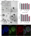
Fig. 1
Fig. 1. Impact of peptide incorporation on hPSC morphology and function. (A) Representative brightfield micrographs comparing WTC-11 and H9 hPSC cultures across different substrate conditions: Cultrex™, terpolymer alone (T), terpolymer with fibronectin (T + FB), and terpolymer with RGD peptide (T + RGD). Scale bars: 200 μm. (B) Cell proliferation measured as live fold expansion for both cell lines when cultured on 2 : 4 : 94 P400 terpolymer (15 wt%) modified with different bioactive molecules (FB, RGD, RGDS; 6 μg per well). (C) Maintenance of pluripotency assessed by OCT4 expression using flow cytometry under identical culture conditions. Data shown as mean ± SD with n = 5. Statistical significance: *p < 0.0001, **p = 0.081, #p = 0.0383 compared to Cultrex™. (D) Immunofluorescence images of WTC-11 hiPSCs cultured on 2 : 4 : 94 P400 terpolymer scaffolds (15 wt%) demonstrating expression of pluripotency markers OCT-4 (green) and TRA-1-81 (red), with nuclear counterstaining using DAPI (blue). Merged images confirm maintenance of stem cell identity. Scale bars: 100 μm.
Fig. 2
Fig. 2. Gene expression profiles and metabolic activity of WTC-11 iPSCs and H9 hESCs cultured on the terpolymer (2 : 4 : 94 P400) with and without the addition of peptides (fibronectin (FB), RGD) at a concentration of 6 μg per well. (A) Relative expression of OCT4 assessed by qPCR for WTC-11 and H9 cells. (B) Relative expression of SOX2 assessed by qPCR for WTC-11 and H9 cells. (C) Relative expression of NANOG assessed by qPCR for H9 cells. (D) Metabolic potential (stressed OCR and ECAR) measured by Seahorse analysis for WTC-11 cells. (E) Metabolic potential (stressed OCR and ECAR) measured by Seahorse analysis for H9 cells. Cultrex™ was used as the control substrate for all comparisons. Statistical comparison between cell lines under T + RGD condition was performed to evaluate differential responses of iPSCs versus ESCs to the same scaffold modification. Statistical significance: *p < 0.03. Bars represent mean ± STDV (n = 3 for gene expression, n = 5 for metabolic potential assays).
Fig. 3
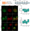
Fig. 3. Maintenance of pluripotency in iPSCs cultured in 3D terpolymer scaffolds with RGD over multiple passages. (A) Schematic representation of the “sandwich” method used for 3D encapsulation of iPSCs in the terpolymer scaffold. (B) A comprehensive multi-passage analysis was performed to evaluate the stability of the 3D culture system supported by the terpolymer scaffold with RGD. (C) Fluorescence microscopy images showing OCT4 expression (green), actin cytoskeleton (red), and merged channels for WTC-11 iPSCs cultured in 3D terpolymer scaffolds across passages 1, 3, and 6. Scale bars represent 100 μm. (D) Flow cytometry quantification of OCT4 expression across passages 1, 2, 4, and 6 (n = 5). (E) Flow cytometry quantification of NANOG expression across the same passages (n = 5).
Fig. 4
Fig. 4. Maintenance of pluripotency and metabolic activity of hPSCs in 3D terpolymer culture. (A) Live fold expansion of iPSCs (WTC-11) and hESCs (H9) encapsulated in 2 : 4 : 94 P400 terpolymer (T) at 15 wt% compared to VitroGel™ commercial synthetic matrix control, with and without addition of fibronectin (T + FB), RGD peptide (T + RGD), or vitronectin (T + VN). (B) Flow cytometry analysis showing percentage of cells expressing pluripotency markers OCT4 and (C) NANOG, across different culture conditions. (D) Relative gene expression of OCT4 and (E) SOX2 assessed by qPCR for WTC-11 and H9 cells. (F) Relative expression of NANOG assessed by qPCR for H9 cells. (G) Metabolic potential measured by Seahorse analysis showing stressed oxygen consumption rate (OCR) and extracellular acidification rate (ECAR) for WTC-11 and (H) H9 cells, respectively. Bars represent mean ± SD (n = 5 for all analyses except gene expression where n = 3). Statistical significance: *p < 0.05, **p = 0.0123, ***p = 0.0004.
Fig. 5
Fig. 5. Encapsulation in terpolymer scaffolds promotes hPSC aggregation and differentiation. (A) Schematic representation of the differentiation protocol from hiPSCs/hESCs into cardiomyocytes within BME and synthetic scaffolds. (B) Representative confocal fluorescence images showing expression of cardiac-specific markers: cardiac Troponin T (cTnT, green), cardiac Troponin I (cTnI, orange), and merged channels in WTC-11 iPSC-derived cardiomyocytes encapsulated in the terpolymer scaffold. Scale bars: 100 μm. (C) Flow cytometry quantification of cTnT and (D) cTnI expression for WTC-11 and H9-derived cardiomyocytes after 15 days of differentiation compared to VitroGel™ (n = 5). (E) Relative gene expression analysis by qPCR of cardiac differentiation markers TNNT2, CACNA1c, and MYH6 for (E) WTC-11 and (F) H9 cell lines cultured in 3D terpolymer scaffolds (n = 3). Data shown as mean ± SD. Statistical significance: *p < 0.05, **p < 0.001.
Fig. 6
Fig. 6. Effect of scaffold stiffness on iPSC proliferation and differentiation into cardiomyocytes. (A) Live fold expansion rates of WTC-11 iPSCs cultured on terpolymer scaffolds with varying stiffness values and control substrates. (B) Expression of pluripotency markers OCT4 and NANOG in iPSCs across different terpolymer stiffness conditions. (C) Percentage of cells expressing cTnT following differentiation on various terpolymer formulations. (D) Percentage of cells expressing cTnI across identical culture conditions. Statistical significance indicated as: ns = not significant. Data represents mean ± SD, n = 4.

References

    1. Peischard S. Piccini I. Strutz-Seebohm N. Greber B. Seebohm G. From iPSC towards cardiac tissue—a road under construction. Pflugers Arch. 2017;469:1233–1243. - PMC - PubMed
    1. Macqueen L. A. et al. , A tissue-engineered scale model of the heart ventricle. Nat. Biomed. Eng. 2018;2:930–941. - PMC - PubMed
    1. Depalma S. J. Davidson C. D. Stis A. E. Helms A. S. Baker B. M. Microenvironmental determinants of organized iPSC-cardiomyocyte tissues on synthetic fibrous matrices. Biomater. Sci. 2021;9:93–107. - PMC - PubMed
    1. Le M. N. T. Hasegawa K. Expansion culture of human pluripotent stem cells and production of cardiomyocytes. Bioengineering. 2019;6:48. - PMC - PubMed
    1. Keung A. J. Dong M. Schaffer D. V. Kumar S. Pan-neuronal maturation but not neuronal subtype differentiation of adult neural stem cells is mechanosensitive. Sci. Rep. 2013;3:1817. - PMC - PubMed

LinkOut - more resources