. 2025 Oct;38(10):e70126.
doi: 10.1002/nbm.70126.

Accelerated Navigator for Rapid ∆B0 Field Mapping for Real-Time Shimming and Motion Correction of Human Brain MRI

Affiliations

Accelerated Navigator for Rapid ∆B0 Field Mapping for Real-Time Shimming and Motion Correction of Human Brain MRI

Nutandev Bikkamane Jayadev et al. NMR Biomed. 2025 Oct.

Abstract

∆B0 shim optimization performed at the beginning of an MR scan is unable to correct for ∆B0 field inhomogeneities caused by patient motion or hardware instability during scans. Navigator-based methods have been demonstrated previously to be effective for motion and shim correction. The purpose of this work was to accelerate volumetric navigators to allow fast acquisition of the parent navigated sequence with short real-time feedback time and high spatial resolution of the ∆B0 field mapping. A GRAPPA-accelerated 3D dual-echo EPI vNav was implemented on a 3 T Prisma MRI scanner. Testing was performed on an anthropomorphic head phantom and 11 human participants. vNav-derived ∆B0 field maps with various spatial resolutions were compared to Cartesian-encoded gold-standard 3D gradient-echo ∆B0 field mapping. ∆B0 shimming was evaluated for the scanner's spherical harmonics shims and a custom-made AC/DC RF-receive/∆B0-shim array. The performance of dual-echo and single-echo accelerated navigators was compared for tracking and updating ∆B0 field maps during motion. Real-time motion and shim corrections for 2D MRI and 3D MRSI sequences were assessed in vivo with controlled head movement. Up to 8-fold acceleration of volumetric navigators (vNavs) significantly reduced geometric distortions and signal dropouts near air-tissue interfaces and metal implants. Acceleration allowed a flexible tradeoff between spatial resolution (2.5-7.5 mm) and acquisition time (242-1302 ms). Notably, accelerated high-resolution (5 mm) vNav was faster (378 ms) than unaccelerated low-resolution (7.5 mm) vNav (700 ms) and showed better agreement with 3D-GRE ∆B0 field mapping with 5.5 Hz RMSE, 1 Hz bias, and [-10%, +10%] confidence interval. Accelerated vNavs improved 3D MRSI and 2D MRI in real-time motion and shim correction applications. Advanced shimming with spherical harmonic and shim array showed superior ΔB0 correction, especially with joint shim optimization. GRAPPA-accelerated vNavs provide fast, robust, and high-quality ∆B0 field mapping and shimming over the whole-brain. The accelerated vNavs enable rapid correction of ∆B0 field inhomogeneities and faster acquisition of the navigated parent sequence. This methodology can be used for real-time motion and shim correction to enhance data quality in various MRI applications.

Keywords: GRAPPA acceleration; magnetic resonance spectroscopic imaging (MRSI); motion correction; multi‐coil shim array; real‐time shimming; spherical harmonic shims; volumetric navigators (vNavs); ΔB₀ field mapping.


Figures

FIGURE 1
FIGURE 1
Navigator acquisition and ∆B0 field mapping/shimming pipeline. (A) Pulse sequence for GRAPPA dual‐echo EPI volume navigator. (B) Channel‐wise data is acquired and processed for combined phase difference, phase unwrapping, brain masking, computing ∆B0 field maps, and calculating shim currents for 2SH, multi‐coil and joint optimization. (C) Uploading of the multi‐coil shim currents to the 32‐channel AC/DC coil.
FIGURE 2
FIGURE 2
Magnitude and ∆B0 field maps from a healthy human volunteer. Gold‐standard 3D‐GRE and EPI‐vNavs (with four resolutions and two accelerations) are shown at their acquired resolution. Respective protocol parameters are tabulated below. The GRAPPA acceleration denotes in‐plane and partition (in‐plane × partition) undersampling factors. The total acquisition time for accelerated sequences (TA*) excludes the duration of the ACS lines.
FIGURE 3
FIGURE 3
Comparison of magnitude and ∆B0 field maps in an anthropomorphic head phantom and a brain tumor patient with metal implants. EPI‐vNavs (with four resolutions and two accelerations) are compared against the gold‐standard 3D‐GRE. The BET brain masks are overlayed on the magnitude images (in green). The low‐resolution vNav images are interpolated to match the 3D‐GRE resolution for statistical comparisons. Corresponding standard deviation for the whole brain volume are shown. ∆B0 map difference, RMSE and histograms for vNavs are shown in comparison to 3D‐GRE. All ∆B0 field maps are measured using the scanner tune‐up shims. The red arrows indicate areas of signal dropout in the unaccelerated vNav, and the blue arrows indicate signal recovery in the same areas with the accelerated vNav.
FIGURE 4
FIGURE 4
Quantitative analysis of ∆B0 field mapping. (A) Histogram, and box plots showing standard deviation and root mean squared error of vNavs with respect to 3D‐GRE. (B) Bland–Altman plots for vNav ∆B0 field maps with respect to 3D‐GRE ∆B0 field maps. All results are based on pooled data from 11 human participants.
FIGURE 5
FIGURE 5
Shimming results comparing ∆B0 field maps from an anthropomorphic head phantom and a healthy human volunteer using all ∆B0 field maps and three shimming methods. The un‐shimmed ∆B0 field maps are acquired with the scanner tune‐up shim for 3D‐GRE and vNav protocols. Optimized ∆B0 shimming was computed for spherical harmonic (2SH), 32‐channel multi‐coil shim array (Multi‐coil) and combined 2SH + Multi‐coil (Joint) hardware. Optimized shims were applied to prospectively acquire ∆B0 field maps using the same 3D‐GRE protocol for all computations. The corresponding standard deviation for the whole brain volume is shown.
FIGURE 6
FIGURE 6
Evaluation of high‐resolution accelerated vNav efficiency for real‐time motion correction in an anthropomorphic head phantom. Top panels show representative 2D‐EPI magnitude images acquired with interleaved vNavs (two resolutions and two acceleration factors) across 30 repeated measurements under four conditions: Static, NoCo (no correction), MoCo (motion correction), and MoShCo (motion and shim correction). Corresponding difference maps depict the relative change between the final (post‐motion) and reference (pre‐motion) images. The phantom was physically rotated and translated consistently for all measurements. Bottom panels show box plots of the percentage difference across the 3D volume for the corresponding condition and vNav configuration. Static shows minimal variability, serving as a baseline. NoCo exhibits significant motion‐induced variation, which is substantially reduced with MoCo and further improved with MoShCo. Red markers indicate mean values; whiskers denote variability.
FIGURE 7
FIGURE 7
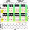
Effect of real‐time motion correction on 2D EPI images using interleaved vNav48PAT4x2 with controlled head movement in a healthy volunteer. The top row shows results with motion correction (Motion MoCo), the middle row shows motion with no correction (Motion NoCo), and motion plots (rotation—Rx,y,z and translation—Tx,y,z) plots are shown at the bottom. Magnitude images and field map relative differences are calculated between the reference (before motion) and final (after motion).
FIGURE 8
FIGURE 8
Effect of real‐time motion and shim correction on 3D MRSI using interleaved vNav48PAT4x2 and an AC/DC shim array under controlled head movement in a healthy volunteer. The figure compares conditions: resting (Rest), head movement with real‐time motion and shim correction (Motion MoShCo), head movement without correction (Motion NoCo), and head movement with only motion correction (Motion MoCo). Metabolic maps of total N‐acetyl‐aspartate (NAA), creatine (Cre), and choline (Cho) are presented, along with spectral linewidth (FWHM) and signal‐to‐noise ratio (SNR) maps. Relative differences are calculated between the static maps as reference and the maps of the three motion experiments. At the bottom, spectra from selected voxels (indicated by arrows on FWHM maps) and motion tracking plots from the navigator are displayed.
FIGURE 9
FIGURE 9
Evaluation of single‐echo ∆B0 field mapping under static and chin‐up motion conditions using an anthropomorphic head phantom with vNav48PAT1 and vNav48PAT4x2 navigators. For a representative slice, single‐echo ∆B0 field maps are compared to dual‐echo ∆B0 field maps for the initial head position (Ref) and the final head position (Final) without doing shim‐update (Left panel) and with the shim‐update (Right panel). Histograms of the bottom show the distribution of ∆B0 over the entire 3D brain volume. The motion plots and shimming coefficients are shown in Figure 10.
FIGURE 10
FIGURE 10
Quantitative evaluation of single‐echo‐derived ΔB₀ field mapping corresponding to the data shown in Figure 9. (A) Mean ΔB₀ difference between single‐echo and dual‐echo field maps across 30 dynamic time‐series measurements for vNav48PAT1 (green) and vNav48PAT4x2 (purple), under static (top) and moved (bottom) conditions, without (left) and with (right) simulated shim correction. (B) Motion parameters estimated from PACE tracking for vNav48PAT1 (left) and vNav48PAT4x2 (right), indicating a ~ 10 mm chin‐up translation (Tz) introduced between measurements 10–15. (C) Simulated shim coefficients (frequency and first‐order X, Y, and Z terms) derived from single‐echo (dashed lines) and dual‐echo (solid lines) ΔB₀ field maps, showing both static (dark blue/light blue) and motion (red/orange) conditions for vNav48PAT1 and vNav48PAT4x2.

Similar articles

References

    1. Liu J., de Zwart J. A., van Gelderen P., Murphy‐Boesch J., and Duyn J. H., “Effect of Head Motion on MRI B0 Field Distribution,” Magnetic Resonance in Medicine 80, no. 6 (2018): 2538–2548, 10.1002/mrm.27339. - DOI - PMC - PubMed
    1. Van de Moortele P., Pfeuffer J., Glover G. H., Ugurbil K., and Hu X., “Respiration‐induced B 0 fluctuations and their spatial distribution in the human brain at 7 Tesla,” Magnetic Resonance in Medicine 47, no. 5 (2002): 888–895, 10.1002/mrm.10145. - DOI - PubMed
    1. Zhou J., Hagberg G. E., Aghaeifar A., Bause J., Zaitsev M., and Scheffler K., “Prediction of Motion Induced Magnetic Fields for Human Brain MRI at 3 T,” Magma 36, no. 5 (2023): 797–813, 10.1007/s10334-023-01076-0. - DOI - PMC - PubMed
    1. Durand E., van de Moortele P., Pachot‐Clouard M., and Le Bihan D., “Artifact Due to B 0 Fluctuations in fMRI: Correction Using the k‐ Space Central Line,” Magnetic Resonance in Medicine 46, no. 1 (2001): 198–201, 10.1002/mrm.1177. - DOI - PubMed
    1. Wezel J., Boer V. O., van der Velden T. A., et al., “A Comparison of Navigators, Snap‐Shot Field Monitoring, and Probe‐Based Field Model Training for Correcting B 0‐Induced Artifacts in ‐Weighted Images at 7 T,” Magnetic Resonance in Medicine 78, no. 4 (2017): 1373–1382, 10.1002/mrm.26524. - DOI - PubMed

LinkOut - more resources