Review
. 2025 Sep 4;23(1):36.
doi: 10.1007/s11914-025-00929-y.

Pathophysiology of Femoral Fractures in Hypophosphatasia

Affiliations
Review

Pathophysiology of Femoral Fractures in Hypophosphatasia

Fabiana G A Tabegna et al. Curr Osteoporos Rep. .

Abstract

Purpose of review: In this review, we will examine the pathophysiology, anatomy, biochemistry, and genotype-phenotype correlation of femoral fractures in adult hypophosphatasia.

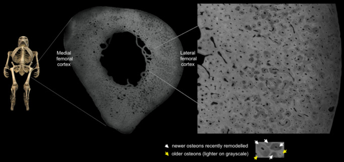
Recent findings: Hypophosphatasia (HPP) is a rare genetic disease characterized by low activity of tissue-nonspecific alkaline phosphatase (TNAP). The disease presents a broad spectrum of clinical manifestations primarily determined by the degree of residual TNAP activity. Adults with HPP of moderate clinical severity may present with spontaneous femoral fractures that are like the atypical femoral fractures (AFF) of long-term bisphosphonates users. In this review, we will focus on the paradox that while HPP can cause biopsy-proven osteomalacia (pathologically impaired bone mineralisation), the spontaneous femoral fractures that characterise adult HPP do not exhibit typical osteomalacia features. Instead, they resemble the femoral fractures that occur in other diseases such as osteopetrosis where bone becomes excessively dense, brittle and highly mineralised due to osteoclast dysfunction. This review examines the key aspects of the pathophysiology of femoral fractures in adults with HPP, offering new insights into the role of anatomical, molecular and biochemical bone abnormalities that characterise the disease. Further investigations of HPP patients with femoral fracture are needed to examine the nanoscale crystal structure of the bone and to study abnormalities in fracture healing and bone resorption.

Keywords: Bone mineralization; Femoral fractures; Hypophosphatasia; Osteomalacia; Osteopetrosis; TNAP.


Conflict of interest statement

Declarations. Ethics Approval and Consent to Participate: The patients described in this review were identified using the Cambridge Outpatient Bone Registry (COBRA), as part of the East Anglian Rare Bone Network (ErBON) and gave informed consent for publication. The study “Characterisation of the mineral content of bone in hypophosphatasia” was approved by the Research Ethics Committee and HRA 22/HRA/0246. Competing Interests: The authors have no competing interests to declare that are relevant to the content of this article.

Figures

Fig. 1
Fig. 1
Forces in the femur during gait. Dark blue arrows. Classical medial femoral ‘Pseudofracture’ or ‘Looser zone’ in cortical bone in a 48-year-old male patient with severe vitamin D deficiency osteomalacia (but not HPP). The fracture line of osteomalacia typically occurs at a zone of high compression (during gait) in the medial femur. Such fractures are painful but rarely complete. Dark red arrows. Although unaffected in this patients’ femur, the red arrows indicate the typical location of highest tension (during gait) in a femur. This lateral cortical site is the stereotypical location of ‘brittle’ fractures which frequently give way to require surgical nailing in hypophosphatasia, genetic osteopetrosis and rarely in long term bisphosphonate users
Fig. 2
Fig. 2
Classical lateral cortical bone fractures in HPP (a) CT scout view (left, main panel) of a 45-year-old female HPP patient whose first presentation was with a spontaneous ‘tensile’ fracture through her dense, thickened right femoral cortex. She was also found to have a left femoral endosteal thickening (arrowed). This endosteal expansion/callus seems widespread in this type of HPP fracture and is noted to be different to the periosteal ‘beaking’ of bisphosphonate-associated fractures. (b) Closer investigation of the left femoral cortex with oblique parasagittal (upper right) and axial (lower right) CT reconstructions showed an impending break with a large plateau of disconnected lateral femoral cortex. White arrows also show the extent of the macroscopically visible tensile fracture lines. Genetic analysis revealed compound heterozygosity at the ALPL gene (NM_000478:5: c.340G > A p.(Ala114Thr) and c.1363G > A p.(Gly455Ser))
Fig. 3
Fig. 3
Adapted from Global hypophosphatasia registry data. Almost half (47%) of adult HPP patients with > 1 ALPL variant sustained subtrochanteric femoral shaft fractures compared with only 8% of those with a single ALPL variant. The femoral shaft was the only fracture type that showed genotype-phenotype interaction [28]
Fig. 4
Fig. 4
The normal lateral subtrochanteric femoral cortex is a site of extensive new osteonal remodelling/ resorption throughout life in response to tensile microdamage. A polished, contact femoral microradiograph of a male aged < 25 years with permission from C.D.L.T, courtesy of the Melbourne Femur collection. The extensive newer osteons are darker grey compared to the established, lighter grey older osteons [59]
Fig. 5
Fig. 5
Toluidine blue stained undecalcified section of the periosteal surface of an iliac bone biopsy from a patient with hypophosphatasia. She has compound heterozygous mutations in ALPL and was admitted with bilateral, spontaneous femur fractures, like the patient in Fig. 1. Multinucleated osteoclasts are present, adjacent to recently resorbed mineralised bone (white arrows). Howship’s lacunae are excessively ‘scalloped’ or ‘jagged’ at the edges which has been noted previously in HPP bone. There is normal bone cell morphology, including osteocytes in the lacunocanalicular network (black arrow). A 6-layer composite Z-stacked image using 40x lens magnification with crossed polarising filters to highlight bone tissue
Fig. 6
Fig. 6
Multisystem involvement in HPP. TNAP: tissue-nonspecific alkaline phosphatase, PLP: pyridoxal 5’-phosphate, PPi: inorganic pyrophosphate, PEA: phosphoethanolamine. Adapted from Conti et al., 2017 [4]

References

    1. Weiss MJ, Ray K, Henthorn PS, Lamb B, Kadesch T, Harris H. ‘Structure of the human liver/bone/kidney alkaline phosphatase gene’. J Biol Chem, 263, 24, pp. 12002–10 PMID– 3165380, 1988. - PubMed
    1. Whyte MP. Hypophosphatasia: an overview for 2017. Bone. 2017;102:15–25. 10.1016/j.bone.2017.02.011. - PubMed
    1. Fallon MD, Teitelbaum SL, Weinstein RS, Goldfischer S, Brown DM, Whyte MP. Hypophosphatasia: clinicopathologic comparison of the infantile, childhood, and adult forms. Med (United States). 1984;63(1). 10.1097/00005792-198401000-00002. - PubMed
    1. Conti F. ‘Hypophosphatasia: clinical manifestation and burden of disease in adult patients’, Clinical Cases in Mineral and Bone Metabolism, vol. 14, no. 2, 2017, 10.11138/ccmbm/2017.14.1.230 - PMC - PubMed
    1. Berkseth KE, Tebben PJ, Drake MT, Hefferan TE, Jewison DE, Wermers RA. ‘Clinical spectrum of hypophosphatasia diagnosed in adults’, Bone, vol. 54, no. 1, pp. 21–27, 2013, 10.1016/j.bone.2013.01.024 - PubMed

MeSH terms

Substances

Supplementary concepts